- PDB-6p4w: XPB helicase in a complex with truncated Bax1 from Sulfurisphaera... -
+
Open data
ID or keywords:
Loading...
-
Basic information
Entry
Database: PDB / ID: 6p4w
Title
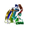
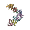
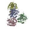
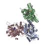
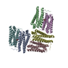
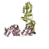
XPB helicase in a complex with truncated Bax1 from Sulfurisphaera tokodaii at 2.96 Angstrom resolution
Components
DNA-dependent ATPase XPBII
Endonuclease Bax1
Keywords
HYDROLASE/DNA BINDING PROTEIN / Helicase / endonuclease / HYDROLASE / HYDROLASE-DNA BINDING PROTEIN complex
Function / homology
Function and homology information
3'-5' DNA helicase activity / DNA 3'-5' helicase / endonuclease activity / Hydrolases; Acting on ester bonds / DNA repair / hydrolase activity / DNA binding / ATP binding Similarity search - Function
Protein of unknown function DUF790, endonuclease-like / Protein of unknown function (DUF790) / Xeroderma pigmentosum group B helicase, damage recognition domain / Xeroderma pigmentosum group B helicase damage recognition domain / Restriction Endonuclease - #30 / ERCC3/RAD25/XPB helicase, C-terminal domain / : / Restriction Endonuclease / Helicase/UvrB, N-terminal / Type III restriction enzyme, res subunit ...Protein of unknown function DUF790, endonuclease-like / Protein of unknown function (DUF790) / Xeroderma pigmentosum group B helicase, damage recognition domain / Xeroderma pigmentosum group B helicase damage recognition domain / Restriction Endonuclease - #30 / ERCC3/RAD25/XPB helicase, C-terminal domain / : / Restriction Endonuclease / Helicase/UvrB, N-terminal / Type III restriction enzyme, res subunit / Helicase conserved C-terminal domain / helicase superfamily c-terminal domain / Superfamilies 1 and 2 helicase C-terminal domain profile. / Superfamilies 1 and 2 helicase ATP-binding type-1 domain profile. / DEAD-like helicases superfamily / Helicase, C-terminal / Helicase superfamily 1/2, ATP-binding domain / P-loop containing nucleotide triphosphate hydrolases / Rossmann fold / P-loop containing nucleoside triphosphate hydrolase / 3-Layer(aba) Sandwich / Alpha Beta Similarity search - Domain/homology
In the structure databanks used in Yorodumi, some data are registered as the other names, "COVID-19 virus" and "2019-nCoV". Here are the details of the virus and the list of structure data.
Jan 31, 2019. EMDB accession codes are about to change! (news from PDBe EMDB page)
EMDB accession codes are about to change! (news from PDBe EMDB page)
The allocation of 4 digits for EMDB accession codes will soon come to an end. Whilst these codes will remain in use, new EMDB accession codes will include an additional digit and will expand incrementally as the available range of codes is exhausted. The current 4-digit format prefixed with “EMD-” (i.e. EMD-XXXX) will advance to a 5-digit format (i.e. EMD-XXXXX), and so on. It is currently estimated that the 4-digit codes will be depleted around Spring 2019, at which point the 5-digit format will come into force.
The EM Navigator/Yorodumi systems omit the EMD- prefix.
Related info.:Q: What is EMD? / ID/Accession-code notation in Yorodumi/EM Navigator
Yorodumi is a browser for structure data from EMDB, PDB, SASBDB, etc.
This page is also the successor to EM Navigator detail page, and also detail information page/front-end page for Omokage search.
The word "yorodu" (or yorozu) is an old Japanese word meaning "ten thousand". "mi" (miru) is to see.
Related info.:EMDB / PDB / SASBDB / Comparison of 3 databanks / Yorodumi Search / Aug 31, 2016. New EM Navigator & Yorodumi / Yorodumi Papers / Jmol/JSmol / Function and homology information / Changes in new EM Navigator and Yorodumi
 Movie
Movie Controller

 Yorodumi
 Open data
 Basic information
 Components
 Keywords
 Function and homology information
 Sulfurisphaera tokodaii (archaea)
 X-RAY DIFFRACTION /
  Authors
 United States, 1items
  Citation
 Structure visualization
 Downloads & links
 Other downloads


 PDBj
PDBj Assembly
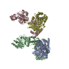


 Mass: 94.971 Da / Num. of mol.: 2 / Source method: obtained synthetically / Formula: PO4
 Mass: 24.305 Da / Num. of mol.: 2 / Source method: obtained synthetically / Formula: Mg
 Mass: 92.094 Da / Num. of mol.: 2 / Source method: obtained synthetically / Formula: C3H8O3
 Mass: 35.453 Da / Num. of mol.: 18 / Source method: obtained synthetically / Formula: Cl
 Sample preparation
 Processing