[English] 日本語
Yorodumi
- PDB-6p4g: Structure of a mammalian small ribosomal subunit in complex with ... -

+
Open data


ID or keywords:

Loading...

-
Basic information

Entry
Database: PDB / ID: 6p4g
TitleStructure of a mammalian small ribosomal subunit in complex with the Israeli Acute Paralysis Virus IRES (Class 1)
Components
  • 18S rRNA
  • IAPV-IRES
  • RACK1
  • eL41
  • eS1
  • eS10
  • eS12
  • eS17
  • eS19
  • eS21
  • eS24
  • eS25
  • eS26
  • eS27
  • eS28
  • eS29
  • eS30
  • eS31
  • eS4
  • eS6
  • eS7
  • eS8
  • uS10
  • uS11
  • uS12
  • uS13
  • uS15
  • uS17
  • uS19
  • uS2
  • uS3
  • uS4
  • uS5
  • uS7
  • uS8
  • uS9
KeywordsRIBOSOME / Israeli Acute Paralysis Virus IRES / IAPV / 40S / small ribosomal subunit
Function / homology
Function and homology information


ribosomal subunit / laminin receptor activity / mammalian oogenesis stage / activation-induced cell death of T cells / positive regulation of signal transduction by p53 class mediator / ubiquitin ligase inhibitor activity / phagocytic cup / 90S preribosome / TOR signaling / endonucleolytic cleavage to generate mature 3'-end of SSU-rRNA from (SSU-rRNA, 5.8S rRNA, LSU-rRNA) ...ribosomal subunit / laminin receptor activity / mammalian oogenesis stage / activation-induced cell death of T cells / positive regulation of signal transduction by p53 class mediator / ubiquitin ligase inhibitor activity / phagocytic cup / 90S preribosome / TOR signaling / endonucleolytic cleavage to generate mature 3'-end of SSU-rRNA from (SSU-rRNA, 5.8S rRNA, LSU-rRNA) / T cell proliferation involved in immune response / erythrocyte development / ribosomal small subunit export from nucleus / translation regulator activity / laminin binding / rough endoplasmic reticulum / endonucleolytic cleavage in ITS1 to separate SSU-rRNA from 5.8S rRNA and LSU-rRNA from tricistronic rRNA transcript (SSU-rRNA, 5.8S rRNA, LSU-rRNA) / gastrulation / MDM2/MDM4 family protein binding / cytosolic ribosome / class I DNA-(apurinic or apyrimidinic site) endonuclease activity / DNA-(apurinic or apyrimidinic site) lyase / rescue of stalled ribosome / maturation of SSU-rRNA from tricistronic rRNA transcript (SSU-rRNA, 5.8S rRNA, LSU-rRNA) / maturation of SSU-rRNA / cellular response to leukemia inhibitory factor / small-subunit processome / protein kinase C binding / positive regulation of apoptotic signaling pathway / positive regulation of protein-containing complex assembly / placenta development / spindle / cytoplasmic ribonucleoprotein granule / modification-dependent protein catabolic process / G1/S transition of mitotic cell cycle / protein tag activity / rRNA processing / ribosomal small subunit biogenesis / rhythmic process / positive regulation of canonical Wnt signaling pathway / small ribosomal subunit rRNA binding / ribosome binding / glucose homeostasis / regulation of translation / ribosomal small subunit assembly / virus receptor activity / small ribosomal subunit / T cell differentiation in thymus / cytosolic small ribosomal subunit / cell body / cytoplasmic translation / perikaryon / mitochondrial inner membrane / postsynaptic density / cell differentiation / rRNA binding / ribosome / protein ubiquitination / structural constituent of ribosome / positive regulation of apoptotic process / positive regulation of protein phosphorylation / ribonucleoprotein complex / translation / cell division / DNA repair / mRNA binding / centrosome / dendrite / ubiquitin protein ligase binding / positive regulation of cell population proliferation / synapse / negative regulation of apoptotic process / nucleolus / apoptotic process / protein kinase binding / perinuclear region of cytoplasm / Golgi apparatus / endoplasmic reticulum / DNA binding / RNA binding / zinc ion binding / membrane / nucleus / metal ion binding / plasma membrane / cytosol / cytoplasm
Similarity search - Function
Ribosomal protein S26 / Ribosomal protein S8e, subdomain / OB fold (Dihydrolipoamide Acetyltransferase, E2P) - #1000 / Ribosomal protein S4, central domain / Phosducin; domain 2 / Ribosomal protein S21 / Alpha-Beta Plaits - #3370 / Hypothetical Cytosolic Protein; Chain: A; / Ribosomal protein S27 / first zn-finger domain of poly(adp-ribose) polymerase-1 ...Ribosomal protein S26 / Ribosomal protein S8e, subdomain / OB fold (Dihydrolipoamide Acetyltransferase, E2P) - #1000 / Ribosomal protein S4, central domain / Phosducin; domain 2 / Ribosomal protein S21 / Alpha-Beta Plaits - #3370 / Hypothetical Cytosolic Protein; Chain: A; / Ribosomal protein S27 / first zn-finger domain of poly(adp-ribose) polymerase-1 / Ribosomal Protein S14/S29 / 30s Ribosomal Protein S14; Chain N / Ribosomal Protein S8; Chain: A, domain 1 - #30 / RNA-binding S4 domain / Ribosomal Protein S7 / Ribosomal protein S7/S5 / Dna Ligase; domain 1 - #10 / Ribosomal protein S11/S14 / Ribosomal protein S10 / Structural Genomics Hypothetical 15.5 Kd Protein In mrcA-pckA Intergenic Region; Chain A / S15/NS1, RNA-binding / SH3 type barrels. - #30 / Ribosomal protein L30/S12 / Double Stranded RNA Binding Domain - #20 / Glucose-6-phosphate isomerase like protein; domain 1 / Ribosomal Protein S8; Chain: A, domain 1 / 40S ribosomal protein SA / Ribosomal Protein S5; domain 2 - #10 / 40S ribosomal protein SA, C-terminal domain / 40S ribosomal protein SA C-terminus / N-terminal domain of TfIIb / Ubiquitin-like protein FUBI / Ribosomal Protein S5; domain 2 / Double Stranded RNA Binding Domain / : / Ribosomal protein S26e signature. / Ribosomal protein L41 / Ribosomal protein L41 / Ribosomal protein S26e / Ribosomal protein S21e, conserved site / Ribosomal protein S26e superfamily / Ribosomal protein S26e / Ribosomal protein S21e signature. / YVTN repeat-like/Quinoprotein amine dehydrogenase / : / Ribosomal protein S12e signature. / Ribosomal protein S12e / Ribosomal protein S5, eukaryotic/archaeal / Ribosomal protein S19e, conserved site / Ribosomal protein S19e signature. / 60s Ribosomal Protein L30; Chain: A; / Ribosomal protein S21e / Ribosomal protein S21e superfamily / Ribosomal protein S21e / Ribosomal protein S2, eukaryotic / Small (40S) ribosomal subunit Asc1/RACK1 / S27a-like superfamily / 40S Ribosomal protein S10 / Ribosomal protein S10, eukaryotic/archaeal / Plectin/S10, N-terminal / Plectin/S10 domain / Ribosomal protein S25 / S25 ribosomal protein / Ribosomal protein S27a / Ribosomal protein S27a / Ribosomal protein S27a / Ribosomal protein S2, eukaryotic/archaeal / Ribosomal protein S8e subdomain, eukaryotes / Ribosomal protein S17e, conserved site / Ribosomal protein S17e signature. / Ribosomal protein S30 / : / Ribosomal protein S30 / 40S ribosomal protein S29/30S ribosomal protein S14 type Z / Ribosomal protein S7e signature. / Ribosomal protein S3, eukaryotic/archaeal / Ribosomal protein S19e / Ribosomal protein S3Ae, conserved site / Ribosomal protein S19e / Ribosomal protein S3Ae signature. / Ribosomal_S19e / Ribosomal protein S27e signature. / Ribosomal protein S4e, N-terminal, conserved site / Ribosomal protein S4e signature. / 40S ribosomal protein S4, C-terminal domain / 40S ribosomal protein S4 C-terminus / Ribosomal protein S8e, conserved site / Ribosomal protein S8e signature. / Ribosomal protein S19A/S15e / Ribosomal protein S17e / Ribosomal protein S17e-like superfamily / Ribosomal S17 / 7 Propeller / Methylamine Dehydrogenase; Chain H / : / Ribosomal protein S6, eukaryotic / Ribosomal S24e conserved site / Ribosomal protein S24e signature. / 40S ribosomal protein S1/3, eukaryotes / Ribosomal protein S4e, N-terminal
Similarity search - Domain/homology
RNA / RNA (> 10) / RNA (> 100) / RNA (> 1000) / Small ribosomal subunit protein eS32 / Small ribosomal subunit protein uS4 / Small ribosomal subunit protein eS12 / Small ribosomal subunit protein uS9 / Small ribosomal subunit protein uS10 / Small ribosomal subunit protein RACK1 ...RNA / RNA (> 10) / RNA (> 100) / RNA (> 1000) / Small ribosomal subunit protein eS32 / Small ribosomal subunit protein uS4 / Small ribosomal subunit protein eS12 / Small ribosomal subunit protein uS9 / Small ribosomal subunit protein uS10 / Small ribosomal subunit protein RACK1 / Ubiquitin-ribosomal protein eS31 fusion protein / Small ribosomal subunit protein uS15 / Small ribosomal subunit protein eS1 / Small ribosomal subunit protein eS7 / Small ribosomal subunit protein uS5 / Small ribosomal subunit protein uS12 / Small ribosomal subunit protein uS11 / 40S ribosomal protein S24 / Ubiquitin-like FUBI-ribosomal protein eS30 fusion protein / Small ribosomal subunit protein eS25 / Small ribosomal subunit protein eS26 / Small ribosomal subunit protein uS7 / Small ribosomal subunit protein uS8 / Small ribosomal subunit protein eS28 / Small ribosomal subunit protein eS8 / Small ribosomal subunit protein eS4 / Small ribosomal subunit protein eS6 / Small ribosomal subunit protein eS21 / Small ribosomal subunit protein eS19 / Small ribosomal subunit protein uS3 / Small ribosomal subunit protein uS13 / Small ribosomal subunit protein eS10 / Small ribosomal subunit protein uS17 / Small ribosomal subunit protein eS17 / Small ribosomal subunit protein uS2 / Small ribosomal subunit protein eS27 / Small ribosomal subunit protein uS19 / Small ribosomal subunit protein uS11 / Small ribosomal subunit protein uS14
Similarity search - Component
Biological speciesIsraeli acute paralysis virus
Oryctolagus cuniculus (rabbit)
MethodELECTRON MICROSCOPY / single particle reconstruction / cryo EM / Resolution: 3.1 Å
AuthorsAcosta-Reyes, F.J. / Neupane, R. / Frank, J. / Fernandez, I.S.
Funding support United States, 1items
OrganizationGrant numberCountry
National Institutes of Health/National Institute of General Medical Sciences (NIH/NIGMS)GM29169 United States
CitationJournal: EMBO J / Year: 2019
Title: The Israeli acute paralysis virus IRES captures host ribosomes by mimicking a ribosomal state with hybrid tRNAs.
Authors: Francisco Acosta-Reyes / Ritam Neupane / Joachim Frank / Israel S Fernández /
Abstract: Colony collapse disorder (CCD) is a multi-faceted syndrome decimating bee populations worldwide, and a group of viruses of the widely distributed Dicistroviridae family have been identified as a ...Colony collapse disorder (CCD) is a multi-faceted syndrome decimating bee populations worldwide, and a group of viruses of the widely distributed Dicistroviridae family have been identified as a causing agent of CCD. This family of viruses employs non-coding RNA sequences, called internal ribosomal entry sites (IRESs), to precisely exploit the host machinery for viral protein production. Using single-particle cryo-electron microscopy (cryo-EM), we have characterized how the IRES of Israeli acute paralysis virus (IAPV) intergenic region captures and redirects translating ribosomes toward viral RNA messages. We reconstituted two in vitro reactions targeting a pre-translocation and a post-translocation state of the IAPV-IRES in the ribosome, allowing us to identify six structures using image processing classification methods. From these, we reconstructed the trajectory of IAPV-IRES from the early small subunit recruitment to the final post-translocated state in the ribosome. An early commitment of IRES/ribosome complexes for global pre-translocation mimicry explains the high efficiency observed for this IRES. Efforts directed toward fighting CCD by targeting the IAPV-IRES using RNA-interference technology are underway, and the structural framework presented here may assist in further refining these approaches.
History
DepositionMay 27, 2019Deposition site: RCSB / Processing site: RCSB
Revision 1.0Sep 18, 2019Provider: repository / Type: Initial release
Revision 1.1Oct 30, 2019Group: Data collection / Database references / Category: citation / citation_author
Item: _citation.country / _citation.journal_abbrev ..._citation.country / _citation.journal_abbrev / _citation.journal_id_ASTM / _citation.journal_id_CSD / _citation.journal_id_ISSN / _citation.page_first / _citation.page_last / _citation.pdbx_database_id_DOI / _citation.pdbx_database_id_PubMed / _citation.title / _citation.year / _citation_author.identifier_ORCID / _citation_author.name
Revision 1.2Nov 13, 2019Group: Database references / Category: citation / Item: _citation.journal_volume
Revision 1.3Jan 8, 2020Group: Author supporting evidence / Category: pdbx_audit_support / Item: _pdbx_audit_support.funding_organization

-
Structure visualization

Movie
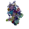
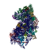
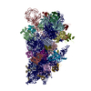
  • Deposited structure unit
  • Imaged by Jmol
  • Download
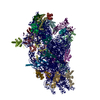
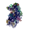
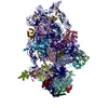
  • Superimposition on EM map
  • EMDB-20248
  • Imaged by UCSF Chimera
  • Download
Movie viewer
Structure viewerMolecule:
MolmilJmol/JSmol

Downloads & links

-
Assembly

Deposited unit
2: 18S rRNA
B: uS2
C: eS1
D: uS5
E: uS3
F: eS4
G: uS7
H: eS6
I: eS7
J: eS8
K: uS4
L: eS10
M: uS17
N: eS12
O: uS15
P: uS11
Q: uS19
R: uS9
S: eS17
T: uS13
U: eS19
V: uS10
W: eS21
X: uS8
Y: uS12
Z: eS24
a: eS25
b: eS26
c: eS27
d: eS28
e: eS29
f: eS30
g: eS31
h: RACK1
n: eL41
1: IAPV-IRES


Theoretical massNumber of molelcules
Total (without water)1,316,41836
Polymers1,316,41836
Non-polymers00
Water00
1


  • Idetical with deposited unit
  • defined by author
  • Evidence: microscopy
TypeNameSymmetry operationNumber
identity operation1_5551

-
Components

-
RNA chain , 2 types, 2 molecules 21

#1: RNA chain 18S rRNA


Mass: 602776.875 Da / Num. of mol.: 1 / Source method: isolated from a natural source / Source: (natural) Oryctolagus cuniculus (rabbit)
#36: RNA chain IAPV-IRES


Mass: 81572.078 Da / Num. of mol.: 1
Source method: isolated from a genetically manipulated source
Source: (gene. exp.) Israeli acute paralysis virus / Production host: synthetic construct (others)

+
Protein , 33 types, 33 molecules BCDEFGHIJKLMNOPQRSTUVWXYZabcde...

#2: Protein uS2 / 37 kDa laminin receptor precursor / 37LRP / 37/67 kDa laminin receptor / LRP/LR / 67 kDa laminin ...37 kDa laminin receptor precursor / 37LRP / 37/67 kDa laminin receptor / LRP/LR / 67 kDa laminin receptor / 67LR / Laminin receptor 1 / LamR / Laminin-binding protein precursor p40 / LBP/p40


Mass: 33003.070 Da / Num. of mol.: 1 / Source method: isolated from a natural source / Source: (natural) Oryctolagus cuniculus (rabbit) / References: UniProt: G1TWL4
#3: Protein eS1


Mass: 30002.061 Da / Num. of mol.: 1 / Source method: isolated from a natural source / Source: (natural) Oryctolagus cuniculus (rabbit) / References: UniProt: G1SS70
#4: Protein uS5


Mass: 27485.209 Da / Num. of mol.: 1 / Source method: isolated from a natural source / Source: (natural) Oryctolagus cuniculus (rabbit) / References: UniProt: G1SWM1
#5: Protein uS3


Mass: 31146.607 Da / Num. of mol.: 1 / Source method: isolated from a natural source / Source: (natural) Oryctolagus cuniculus (rabbit) / References: UniProt: G1TNM3
#6: Protein eS4


Mass: 29658.920 Da / Num. of mol.: 1 / Source method: isolated from a natural source / Source: (natural) Oryctolagus cuniculus (rabbit) / References: UniProt: G1TK17
#7: Protein uS7


Mass: 22913.453 Da / Num. of mol.: 1 / Source method: isolated from a natural source / Source: (natural) Oryctolagus cuniculus (rabbit) / References: UniProt: G1TFM5
#8: Protein eS6


Mass: 28751.906 Da / Num. of mol.: 1 / Source method: isolated from a natural source / Source: (natural) Oryctolagus cuniculus (rabbit) / References: UniProt: G1TM55
#9: Protein eS7


Mass: 22168.914 Da / Num. of mol.: 1 / Source method: isolated from a natural source / Source: (natural) Oryctolagus cuniculus (rabbit) / References: UniProt: G1SVB0
#10: Protein eS8


Mass: 24263.387 Da / Num. of mol.: 1 / Source method: isolated from a natural source / Source: (natural) Oryctolagus cuniculus (rabbit) / References: UniProt: G1TJW1
#11: Protein uS4


Mass: 22641.564 Da / Num. of mol.: 1 / Source method: isolated from a natural source / Source: (natural) Oryctolagus cuniculus (rabbit) / References: UniProt: B7NZS8
#12: Protein eS10


Mass: 17156.865 Da / Num. of mol.: 1 / Source method: isolated from a natural source / Source: (natural) Oryctolagus cuniculus (rabbit) / References: UniProt: G1TPV3
#13: Protein uS17


Mass: 18468.826 Da / Num. of mol.: 1 / Source method: isolated from a natural source / Source: (natural) Oryctolagus cuniculus (rabbit) / References: UniProt: G1TRM4
#14: Protein eS12


Mass: 14538.987 Da / Num. of mol.: 1 / Source method: isolated from a natural source / Source: (natural) Oryctolagus cuniculus (rabbit) / References: UniProt: G1SFR8
#15: Protein uS15


Mass: 17259.389 Da / Num. of mol.: 1 / Source method: isolated from a natural source / Source: (natural) Oryctolagus cuniculus (rabbit) / References: UniProt: G1SP51
#16: Protein uS11


Mass: 16302.772 Da / Num. of mol.: 1 / Source method: isolated from a natural source / Source: (natural) Oryctolagus cuniculus (rabbit) / References: UniProt: G1U472, UniProt: G1T1F0*PLUS
#17: Protein uS19


Mass: 17049.182 Da / Num. of mol.: 1 / Source method: isolated from a natural source / Source: (natural) Oryctolagus cuniculus (rabbit) / References: UniProt: G1U0Q2
#18: Protein uS9


Mass: 19213.465 Da / Num. of mol.: 1 / Source method: isolated from a natural source / Source: (natural) Oryctolagus cuniculus (rabbit) / References: UniProt: G1SGX4
#19: Protein eS17


Mass: 15552.119 Da / Num. of mol.: 1 / Source method: isolated from a natural source / Source: (natural) Oryctolagus cuniculus (rabbit) / References: UniProt: G1TU13
#20: Protein uS13


Mass: 17759.777 Da / Num. of mol.: 1 / Source method: isolated from a natural source / Source: (natural) Oryctolagus cuniculus (rabbit) / References: UniProt: G1TPG3
#21: Protein eS19


Mass: 16235.796 Da / Num. of mol.: 1 / Source method: isolated from a natural source / Source: (natural) Oryctolagus cuniculus (rabbit) / References: UniProt: G1TN62
#22: Protein uS10


Mass: 13398.763 Da / Num. of mol.: 1 / Source method: isolated from a natural source / Source: (natural) Oryctolagus cuniculus (rabbit) / References: UniProt: G1SIZ2
#23: Protein eS21


Mass: 9043.276 Da / Num. of mol.: 1 / Source method: isolated from a natural source / Source: (natural) Oryctolagus cuniculus (rabbit) / References: UniProt: G1TM82
#24: Protein uS8


Mass: 14865.555 Da / Num. of mol.: 1 / Source method: isolated from a natural source / Source: (natural) Oryctolagus cuniculus (rabbit) / References: UniProt: G1TG89
#25: Protein uS12


Mass: 15784.549 Da / Num. of mol.: 1 / Source method: isolated from a natural source / Source: (natural) Oryctolagus cuniculus (rabbit) / References: UniProt: G1SZ47
#26: Protein eS24


Mass: 15548.398 Da / Num. of mol.: 1 / Source method: isolated from a natural source / Source: (natural) Oryctolagus cuniculus (rabbit) / References: UniProt: G1T3D8
#27: Protein eS25


Mass: 13776.224 Da / Num. of mol.: 1 / Source method: isolated from a natural source / Source: (natural) Oryctolagus cuniculus (rabbit) / References: UniProt: G1TDB3
#28: Protein eS26


Mass: 12961.455 Da / Num. of mol.: 1 / Source method: isolated from a natural source / Source: (natural) Oryctolagus cuniculus (rabbit) / References: UniProt: G1TFE8
#29: Protein eS27


Mass: 9480.186 Da / Num. of mol.: 1 / Source method: isolated from a natural source / Source: (natural) Oryctolagus cuniculus (rabbit) / References: UniProt: G1TZ76
#30: Protein eS28


Mass: 7855.052 Da / Num. of mol.: 1 / Source method: isolated from a natural source / Source: (natural) Oryctolagus cuniculus (rabbit) / References: UniProt: G1TIB4
#31: Protein eS29


Mass: 6690.821 Da / Num. of mol.: 1 / Source method: isolated from a natural source / Source: (natural) Oryctolagus cuniculus (rabbit) / References: UniProt: G1U7M4
#32: Protein eS30


Mass: 14498.884 Da / Num. of mol.: 1 / Source method: isolated from a natural source / Source: (natural) Oryctolagus cuniculus (rabbit) / References: UniProt: G1T8A2
#33: Protein eS31


Mass: 18004.041 Da / Num. of mol.: 1 / Source method: isolated from a natural source / Source: (natural) Oryctolagus cuniculus (rabbit) / References: UniProt: G1SK22
#34: Protein RACK1


Mass: 35115.652 Da / Num. of mol.: 1 / Source method: isolated from a natural source / Source: (natural) Oryctolagus cuniculus (rabbit) / References: UniProt: G1SJB4

-
Protein/peptide , 1 types, 1 molecules n

#35: Protein/peptide eL41


Mass: 3473.451 Da / Num. of mol.: 1 / Source method: isolated from a natural source / Source: (natural) Oryctolagus cuniculus (rabbit) / References: UniProt: A0A087WNH4

-
Experimental details

-
Experiment

ExperimentMethod: ELECTRON MICROSCOPY
EM experimentAggregation state: PARTICLE / 3D reconstruction method: single particle reconstruction

-
Sample preparation

ComponentName: Structure of a mammalian small ribosomal subunit in complex with the Israeli Acute Paralysis Virus IRES (Class 1)
Type: RIBOSOME / Entity ID: all / Source: MULTIPLE SOURCES
Molecular weightExperimental value: NO
Source (natural)Organism: Israeli acute paralysis virus
Source (recombinant)Organism: Israeli acute paralysis virus
Buffer solution
IDSpecimen-IDpH
117.5
227.5
Buffer component
IDConc.FormulaBuffer-ID
120 mMTris-HCl1
2100 mMKCl1
38 mMMgCl21
42 mMDTT1
52
Specimen
IDExperiment-IDEmbedding appliedShadowing appliedStaining appliedVitrification appliedDetails
11NONONOYESRibosomal complexes for the pre-translocated state were assembled at 240-390 nM concentration and applied to plasma treated holey carbon grids.
21NONONOYESSame sample and buffer conditions used for the holey carbon grids was used for the holey gold grids.
Specimen support
IDSpecimen-IDDetailsGrid materialGrid typeGrid mesh size (divisions/in.)
11Plasma cleaning for both holey carbon and holey gold grids was done on a Gatan Solarus with Hydrogen (6.4 sccm gas flow) and Oxygen (27.5 sccm gas flow) and 10 W cleaning power.COPPERQuantifoil R2/2
22Plasma cleaning for both holey carbon and holey gold grids was done on a Gatan Solarus with Hydrogen (6.4 sccm gas flow) and Oxygen (27.5 sccm gas flow) and 10 W cleaning power.GOLDQuantifoil, UltrAuFoil, R1.2/1.3300
Vitrification

Chamber temperature: 277.15 K / Cryogen name: ETHANE / Details: Blot force = 3s Wait time = 15s Drain time = 0s Blot time = 2.5 to 3 s / Entry-ID: 6P4G / Humidity: 100 % / Instrument: FEI VITROBOT MARK IV

IDSpecimen-ID
11
22

-
Electron microscopy imaging

Experimental equipment
Model: Tecnai F30 / Image courtesy: FEI Company
MicroscopyModel: FEI TECNAI F30
Electron gunElectron source: FIELD EMISSION GUN / Accelerating voltage: 300 kV / Illumination mode: FLOOD BEAM
Electron lensMode: BRIGHT FIELD / Nominal magnification: 31000 X / Nominal defocus max: 2500 nm / Nominal defocus min: 800 nm / Cs: 2.26 mm
Specimen holderCryogen: NITROGEN
Image recordingAverage exposure time: 8 sec. / Electron dose: 42.1 e/Å2 / Detector mode: COUNTING / Film or detector model: GATAN K2 SUMMIT (4k x 4k) / Num. of real images: 11234
Image scansWidth: 3710 / Height: 3838 / Movie frames/image: 40 / Used frames/image: 1-40

-
Processing

SoftwareName: REFMAC / Version: 5.8.0232 / Classification: refinement
EM software
IDNameCategory
1Gautomatchparticle selection
2Leginonimage acquisition
4GctfCTF correction
9RELIONinitial Euler assignment
10RELIONfinal Euler assignment
11RELIONclassification
CTF correctionType: PHASE FLIPPING AND AMPLITUDE CORRECTION
Particle selectionNum. of particles selected: 1240275
SymmetryPoint symmetry: C1 (asymmetric)
3D reconstructionResolution: 3.1 Å / Resolution method: FSC 0.143 CUT-OFF / Num. of particles: 91056 / Symmetry type: POINT
RefinementResolution: 3.1→255.23 Å / Cor.coef. Fo:Fc: 0.859 / SU B: 18.645 / SU ML: 0.284 / ESU R: 0.417
Stereochemistry target values: MAXIMUM LIKELIHOOD WITH PHASES
Details: HYDROGENS HAVE BEEN ADDED IN THE RIDING POSITIONS
RfactorNum. reflection% reflection
Rwork0.35594 --
obs0.35594 872093 100 %
Solvent computationIon probe radii: 0.8 Å / Shrinkage radii: 0.8 Å / VDW probe radii: 1.2 Å / Solvent model: MASK
Displacement parametersBiso mean: 120.142 Å2
Baniso -1Baniso -2Baniso -3
1-1.35 Å2-0.72 Å2-0.04 Å2
2---0.62 Å2-0.76 Å2
3----0.72 Å2
Refinement stepCycle: 1 / Total: 79338
Refine LS restraints
Refine-IDTypeDev idealDev ideal targetNumber
ELECTRON MICROSCOPYr_bond_refined_d0.0040.01284791
ELECTRON MICROSCOPYr_bond_other_d0.0020.01857778
ELECTRON MICROSCOPYr_angle_refined_deg1.1621.441123575
ELECTRON MICROSCOPYr_angle_other_deg1.2342.059135805
ELECTRON MICROSCOPYr_dihedral_angle_1_deg8.4154821
ELECTRON MICROSCOPYr_dihedral_angle_2_deg26.68619.722144
ELECTRON MICROSCOPYr_dihedral_angle_3_deg13.868157751
ELECTRON MICROSCOPYr_dihedral_angle_4_deg13.62915438
ELECTRON MICROSCOPYr_chiral_restr0.0640.212663
ELECTRON MICROSCOPYr_gen_planes_refined0.0040.0265796
ELECTRON MICROSCOPYr_gen_planes_other0.0020.0218738
ELECTRON MICROSCOPYr_nbd_refined
ELECTRON MICROSCOPYr_nbd_other
ELECTRON MICROSCOPYr_nbtor_refined
ELECTRON MICROSCOPYr_nbtor_other
ELECTRON MICROSCOPYr_xyhbond_nbd_refined
ELECTRON MICROSCOPYr_xyhbond_nbd_other
ELECTRON MICROSCOPYr_metal_ion_refined
ELECTRON MICROSCOPYr_metal_ion_other
ELECTRON MICROSCOPYr_symmetry_vdw_refined
ELECTRON MICROSCOPYr_symmetry_vdw_other
ELECTRON MICROSCOPYr_symmetry_hbond_refined
ELECTRON MICROSCOPYr_symmetry_hbond_other
ELECTRON MICROSCOPYr_symmetry_metal_ion_refined
ELECTRON MICROSCOPYr_symmetry_metal_ion_other
ELECTRON MICROSCOPYr_mcbond_it4.28812.6319395
ELECTRON MICROSCOPYr_mcbond_other4.28712.6319394
ELECTRON MICROSCOPYr_mcangle_it7.26518.93424179
ELECTRON MICROSCOPYr_mcangle_other7.26518.93424180
ELECTRON MICROSCOPYr_scbond_it3.74712.84365396
ELECTRON MICROSCOPYr_scbond_other3.74712.84365395
ELECTRON MICROSCOPYr_scangle_it
ELECTRON MICROSCOPYr_scangle_other6.35519.19399397
ELECTRON MICROSCOPYr_long_range_B_refined10.709100211
ELECTRON MICROSCOPYr_long_range_B_other10.709100212
ELECTRON MICROSCOPYr_rigid_bond_restr
ELECTRON MICROSCOPYr_sphericity_free
ELECTRON MICROSCOPYr_sphericity_bonded
LS refinement shellResolution: 3.1→3.181 Å / Total num. of bins used: 20
RfactorNum. reflection% reflection
Rfree0 0 -
Rwork0.608 64506 -
obs--100 %

+
About Yorodumi

-
News

-
Feb 9, 2022. New format data for meta-information of EMDB entries

New format data for meta-information of EMDB entries

  • Version 3 of the EMDB header file is now the official format.
  • The previous official version 1.9 will be removed from the archive.

Related info.:EMDB header

External links:wwPDB to switch to version 3 of the EMDB data model

-
Aug 12, 2020. Covid-19 info

Covid-19 info

URL: https://pdbj.org/emnavi/covid19.php

New page: Covid-19 featured information page in EM Navigator.

Related info.:Covid-19 info / Mar 5, 2020. Novel coronavirus structure data

+
Mar 5, 2020. Novel coronavirus structure data

Novel coronavirus structure data

Related info.:Yorodumi Speices / Aug 12, 2020. Covid-19 info

External links:COVID-19 featured content - PDBj / Molecule of the Month (242):Coronavirus Proteases

+
Jan 31, 2019. EMDB accession codes are about to change! (news from PDBe EMDB page)

EMDB accession codes are about to change! (news from PDBe EMDB page)

  • The allocation of 4 digits for EMDB accession codes will soon come to an end. Whilst these codes will remain in use, new EMDB accession codes will include an additional digit and will expand incrementally as the available range of codes is exhausted. The current 4-digit format prefixed with “EMD-” (i.e. EMD-XXXX) will advance to a 5-digit format (i.e. EMD-XXXXX), and so on. It is currently estimated that the 4-digit codes will be depleted around Spring 2019, at which point the 5-digit format will come into force.
  • The EM Navigator/Yorodumi systems omit the EMD- prefix.

Related info.:Q: What is EMD? / ID/Accession-code notation in Yorodumi/EM Navigator

External links:EMDB Accession Codes are Changing Soon! / Contact to PDBj

+
Jul 12, 2017. Major update of PDB

Major update of PDB

  • wwPDB released updated PDB data conforming to the new PDBx/mmCIF dictionary.
  • This is a major update changing the version number from 4 to 5, and with Remediation, in which all the entries are updated.
  • In this update, many items about electron microscopy experimental information are reorganized (e.g. em_software).
  • Now, EM Navigator and Yorodumi are based on the updated data.

External links:wwPDB Remediation / Enriched Model Files Conforming to OneDep Data Standards Now Available in the PDB FTP Archive

-
Yorodumi

Thousand views of thousand structures

  • Yorodumi is a browser for structure data from EMDB, PDB, SASBDB, etc.
  • This page is also the successor to EM Navigator detail page, and also detail information page/front-end page for Omokage search.
  • The word "yorodu" (or yorozu) is an old Japanese word meaning "ten thousand". "mi" (miru) is to see.

Related info.:EMDB / PDB / SASBDB / Comparison of 3 databanks / Yorodumi Search / Aug 31, 2016. New EM Navigator & Yorodumi / Yorodumi Papers / Jmol/JSmol / Function and homology information / Changes in new EM Navigator and Yorodumi

Read more