 Movie
Movie Controller

[English] 日本語
 Yorodumi
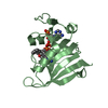
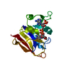
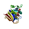
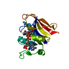
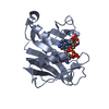
- PDB-6p28: Crystal structure of the MIR domain (aa 337-532) of the S. cerevi... -
+ Open data

- Basic information
| Entry | Database: PDB / ID: 6p28 | ||||||
|---|---|---|---|---|---|---|---|
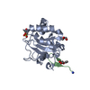
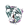
| Title | Crystal structure of the MIR domain (aa 337-532) of the S. cerevisiae mannosyltransferase Pmt2 | ||||||
 Components Components | Dolichyl-phosphate-mannose--protein mannosyltransferase 2 | ||||||
 Keywords Keywords | TRANSFERASE / complex / glycosylation | ||||||
| Function / homology |  Function and homology information Function and homology informationdolichyl-phosphate-mannose-protein mannosyltransferase Pmt1p-Pmt2p dimer complex / dolichyl-phosphate-mannose-protein mannosyltransferase Pmt5p-Pmt2p dimer complex / regulation of endoplasmic reticulum unfolded protein response / dolichyl-phosphate-mannose-protein mannosyltransferase / dolichyl-phosphate-mannose-protein mannosyltransferase activity / fungal-type cell wall biogenesis / protein O-linked glycosylation via mannose / protein exit from endoplasmic reticulum / protein O-linked glycosylation / ERAD pathway ...dolichyl-phosphate-mannose-protein mannosyltransferase Pmt1p-Pmt2p dimer complex / dolichyl-phosphate-mannose-protein mannosyltransferase Pmt5p-Pmt2p dimer complex / regulation of endoplasmic reticulum unfolded protein response / dolichyl-phosphate-mannose-protein mannosyltransferase / dolichyl-phosphate-mannose-protein mannosyltransferase activity / fungal-type cell wall biogenesis / protein O-linked glycosylation via mannose / protein exit from endoplasmic reticulum / protein O-linked glycosylation / ERAD pathway / endoplasmic reticulum membrane / endoplasmic reticulum Similarity search - Function | ||||||
| Biological species |  | ||||||
| Method |  X-RAY DIFFRACTION / SYNCHROTRON / MOLECULAR REPLACEMENT / Resolution: 1.35 Å X-RAY DIFFRACTION / SYNCHROTRON / MOLECULAR REPLACEMENT / Resolution: 1.35 Å | ||||||
 Authors Authors | Bai, L. / Li, H. | ||||||
| Funding support |  United States, 1items United States, 1items
| ||||||
 Citation Citation | Journal: Nat Struct Mol Biol / Year: 2019 Title: Structure of the eukaryotic protein O-mannosyltransferase Pmt1-Pmt2 complex. Authors: Lin Bai / Amanda Kovach / Qinglong You / Alanna Kenny / Huilin Li / Abstract: In eukaryotes, a nascent peptide entering the endoplasmic reticulum (ER) is scanned by two Sec61 translocon-associated large membrane machines for protein N-glycosylation and protein O-mannosylation, ...In eukaryotes, a nascent peptide entering the endoplasmic reticulum (ER) is scanned by two Sec61 translocon-associated large membrane machines for protein N-glycosylation and protein O-mannosylation, respectively. While the structure of the eight-protein oligosaccharyltransferase complex has been determined recently, the structures of mannosyltransferases of the PMT family, which are an integral part of ER protein homeostasis, are still unknown. Here we report cryo-EM structures of the Saccharomyces cerevisiae Pmt1-Pmt2 complex bound to a donor and an acceptor peptide at 3.2-Å resolution, showing that each subunit contains 11 transmembrane helices and a lumenal β-trefoil fold termed the MIR domain. The structures reveal the substrate recognition model and confirm an inverting mannosyl-transferring reaction mechanism by the enzyme complex. Furthermore, we found that the transmembrane domains of Pmt1 and Pmt2 share a structural fold with the catalytic subunits of oligosaccharyltransferases, confirming a previously proposed evolutionary relationship between protein O-mannosylation and protein N-glycosylation. | ||||||
| History |
|
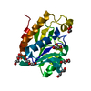
- Structure visualization
| Structure viewer | Molecule: MolmilJmol/JSmol |
|---|
- Downloads & links
-Download
| PDBx/mmCIF format | 6p28.cif.gz | 57.4 KB | Display | PDBx/mmCIF format |
|---|---|---|---|---|
| PDB format | pdb6p28.ent.gz | 40.1 KB | Display | PDB format |
| PDBx/mmJSON format | 6p28.json.gz | Tree view | PDBx/mmJSON format | |
| Others |  Other downloads Other downloads |
-Validation report
| Arichive directory | https://data.pdbj.org/pub/pdb/validation_reports/p2/6p28ftp://data.pdbj.org/pub/pdb/validation_reports/p2/6p28 | HTTPS FTP |
|---|
-Related structure data
| Related structure data |  6p25SC  6p2rC  20237 C: citing same article ( S: Starting model for refinement |
|---|---|
| Similar structure data |


-Links
 PDBj
PDBj- Assembly
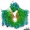
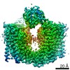
| Deposited unit | 
| ||||||||
|---|---|---|---|---|---|---|---|---|---|
| 1 |
| ||||||||
| Unit cell |
|
-Components
| #1: Protein | Mass: 22498.826 Da / Num. of mol.: 1 / Fragment: MIR domain (UNP residues 337-532) Source method: isolated from a genetically manipulated source Source: (gene. exp.) Gene: PMT2, FUN25, YAL023C / Production host:  References: UniProt: P31382, dolichyl-phosphate-mannose-protein mannosyltransferase |
|---|---|
| #2: Water | ChemComp-HOH /  Mass: 18.015 Da / Num. of mol.: 191 / Source method: isolated from a natural source / Formula: H2O Mass: 18.015 Da / Num. of mol.: 191 / Source method: isolated from a natural source / Formula: H2O |
| Has protein modification | Y |
-Experimental details
-Experiment
| Experiment | Method: X-RAY DIFFRACTION / Number of used crystals: 1 |
|---|
- Sample preparation
| Crystal | Density Matthews: 2.09 Å3/Da / Density % sol: 41.06 % |
|---|---|
| Crystal grow | Temperature: 293 K / Method: vapor diffusion, sitting drop / Details: 25% PEG20000 MME |
-Data collection
| Diffraction | Mean temperature: 100 K / Serial crystal experiment: N |
|---|---|
| Diffraction source | Source: SYNCHROTRON / Site: APS / Beamline: 21-ID-D / Wavelength: 1.078 Å |
| Detector | Type: DECTRIS EIGER X 9M / Detector: PIXEL / Date: Oct 8, 2018 |
| Radiation | Monochromator: Si(111) / Protocol: SINGLE WAVELENGTH / Monochromatic (M) / Laue (L): M / Scattering type: x-ray |
| Radiation wavelength | Wavelength: 1.078 Å / Relative weight: 1 |
| Reflection | Resolution: 1.35→37.65 Å / Num. obs: 40003 / % possible obs: 96.7 % / Redundancy: 3.5 % / Rmerge(I) obs: 0.134 / Net I/σ(I): 13.4 |
| Reflection shell | Resolution: 1.35→1.38 Å / Rmerge(I) obs: 0.023 / Num. unique obs: 17096 |
- Processing
| Software |
| ||||||||||||||||||||||||||||||||||||||||||||||||||||||||||||||||||||||||||||||||||||||||||||||||||||||||||||||||||||||||||||||||||||||||||||||||||||||||||||||||||||||||||||||||||||||
|---|---|---|---|---|---|---|---|---|---|---|---|---|---|---|---|---|---|---|---|---|---|---|---|---|---|---|---|---|---|---|---|---|---|---|---|---|---|---|---|---|---|---|---|---|---|---|---|---|---|---|---|---|---|---|---|---|---|---|---|---|---|---|---|---|---|---|---|---|---|---|---|---|---|---|---|---|---|---|---|---|---|---|---|---|---|---|---|---|---|---|---|---|---|---|---|---|---|---|---|---|---|---|---|---|---|---|---|---|---|---|---|---|---|---|---|---|---|---|---|---|---|---|---|---|---|---|---|---|---|---|---|---|---|---|---|---|---|---|---|---|---|---|---|---|---|---|---|---|---|---|---|---|---|---|---|---|---|---|---|---|---|---|---|---|---|---|---|---|---|---|---|---|---|---|---|---|---|---|---|---|---|---|---|
| Refinement | Method to determine structure: MOLECULAR REPLACEMENT Starting model: PDB entry 6P25 Resolution: 1.35→37.65 Å / Cor.coef. Fo:Fc: 0.972 / Cor.coef. Fo:Fc free: 0.962 / SU B: 0.924 / SU ML: 0.038 / Cross valid method: THROUGHOUT / ESU R: 0.057 / ESU R Free: 0.06 / Details: HYDROGENS HAVE BEEN ADDED IN THE RIDING POSITIONS
| ||||||||||||||||||||||||||||||||||||||||||||||||||||||||||||||||||||||||||||||||||||||||||||||||||||||||||||||||||||||||||||||||||||||||||||||||||||||||||||||||||||||||||||||||||||||
| Solvent computation | Ion probe radii: 0.8 Å / Shrinkage radii: 0.8 Å / VDW probe radii: 1.2 Å | ||||||||||||||||||||||||||||||||||||||||||||||||||||||||||||||||||||||||||||||||||||||||||||||||||||||||||||||||||||||||||||||||||||||||||||||||||||||||||||||||||||||||||||||||||||||
| Displacement parameters | Biso mean: 19.885 Å2
| ||||||||||||||||||||||||||||||||||||||||||||||||||||||||||||||||||||||||||||||||||||||||||||||||||||||||||||||||||||||||||||||||||||||||||||||||||||||||||||||||||||||||||||||||||||||
| Refinement step | Cycle: 1 / Resolution: 1.35→37.65 Å
| ||||||||||||||||||||||||||||||||||||||||||||||||||||||||||||||||||||||||||||||||||||||||||||||||||||||||||||||||||||||||||||||||||||||||||||||||||||||||||||||||||||||||||||||||||||||
| Refine LS restraints |
|