 Movie
Movie Controller


+ Open data

- Basic information
| Entry | Database: PDB / ID: 6ol3 | ||||||
|---|---|---|---|---|---|---|---|
| Title | Crystal structure of an adenovirus virus-associated RNA | ||||||
 Components Components | Adenovirus Virus-Associated (VA) RNA I apical and central domains | ||||||
 Keywords Keywords | RNA / noncoding RNA / viral RNA | ||||||
| Function / homology | : / RNA / RNA (> 10) / RNA (> 100) Function and homology information Function and homology information | ||||||
| Biological species |   Human adenovirus 2 Human adenovirus 2 | ||||||
| Method |  X-RAY DIFFRACTION / SYNCHROTRON / SAD / Resolution: 2.74 Å X-RAY DIFFRACTION / SYNCHROTRON / SAD / Resolution: 2.74 Å | ||||||
 Authors Authors | Hood, I.V. / Gordon, J.M. / Bou-Nader, C. / Henderson, F.V. / Bahmanjah, S. / Zhang, J. | ||||||
| Funding support |  United States, 1items United States, 1items
| ||||||
 Citation Citation | Journal: Nat Commun / Year: 2019 Title: Crystal structure of an adenovirus virus-associated RNA. Authors: Hood, I.V. / Gordon, J.M. / Bou-Nader, C. / Henderson, F.E. / Bahmanjah, S. / Zhang, J. | ||||||
| History |
|
- Structure visualization
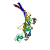
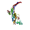
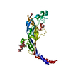
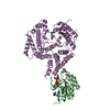
| Structure viewer | Molecule: MolmilJmol/JSmol |
|---|
- Downloads & links
-Download
| PDBx/mmCIF format | 6ol3.cif.gz | 156.9 KB | Display | PDBx/mmCIF format |
|---|---|---|---|---|
| PDB format | pdb6ol3.ent.gz | 125.3 KB | Display | PDB format |
| PDBx/mmJSON format | 6ol3.json.gz | Tree view | PDBx/mmJSON format | |
| Others |  Other downloads Other downloads |
-Validation report
| Arichive directory | https://data.pdbj.org/pub/pdb/validation_reports/ol/6ol3ftp://data.pdbj.org/pub/pdb/validation_reports/ol/6ol3 | HTTPS FTP |
|---|
-Related structure data
| Similar structure data |
|---|


-Links
 PDBj
PDBj


- Assembly
| Deposited unit | 
| ||||||||
|---|---|---|---|---|---|---|---|---|---|
| 1 |
| ||||||||
| Unit cell |
|
-Components
| #1: RNA chain | Mass: 36122.469 Da / Num. of mol.: 1 / Source method: obtained synthetically / Source: (synth.) Human adenovirus 2 |
|---|---|
| #2: Chemical | ChemComp-K /   Mass: 39.098 Da / Num. of mol.: 1 / Source method: obtained synthetically / Formula: K Mass: 39.098 Da / Num. of mol.: 1 / Source method: obtained synthetically / Formula: K |
| #3: Water | ChemComp-HOH /  Mass: 18.015 Da / Num. of mol.: 9 / Source method: isolated from a natural source / Formula: H2O Mass: 18.015 Da / Num. of mol.: 9 / Source method: isolated from a natural source / Formula: H2O |
-Experimental details
-Experiment
| Experiment | Method: X-RAY DIFFRACTION / Number of used crystals: 1 |
|---|
- Sample preparation
| Crystal | Density Matthews: 4.56 Å3/Da / Density % sol: 73.05 % / Description: obelisk crystals |
|---|---|
| Crystal grow | Temperature: 293 K / Method: vapor diffusion, hanging drop / pH: 7 Details: 50 mM sodium cacodylate (pH 6.5) 28-30% 2-methyl-2,4-pentanediol (MPD) |
-Data collection
| Diffraction | Mean temperature: 100 K / Serial crystal experiment: N |
|---|---|
| Diffraction source | Source: SYNCHROTRON / Site: APS / Beamline: 22-ID / Wavelength: 1 Å |
| Detector | Type: RAYONIX MX300-HS / Detector: CCD / Date: Aug 22, 2016 |
| Radiation | Monochromator: MD2 / Protocol: SINGLE WAVELENGTH / Monochromatic (M) / Laue (L): M / Scattering type: x-ray |
| Radiation wavelength | Wavelength: 1 Å / Relative weight: 1 |
| Reflection | Resolution: 2.74→40 Å / Num. obs: 18246 / % possible obs: 99.9 % / Observed criterion σ(F): 0.82 / Redundancy: 8.2 % Data reduction details: EDS server used different resolution to calculate completeness. The higher Rmerge is partly due to the higher redundancy Rmerge(I) obs: 0.152 / Net I/σ(I): 12.2 |
| Reflection shell | Resolution: 2.74→2.81 Å / Redundancy: 8.2 % / Rmerge(I) obs: 3.642 / Mean I/σ(I) obs: 0.82 / Num. unique obs: 1310 / CC1/2: 0.34 / Rrim(I) all: 3.884 / % possible all: 100 |
- Processing
| Software |
| ||||||||||||||||||||||||||||||||||||||||||||||||||||||||||||||||||||||||||||||||||||||||||||||||||||||||||||||||||||||||||||||||||||||||||||
|---|---|---|---|---|---|---|---|---|---|---|---|---|---|---|---|---|---|---|---|---|---|---|---|---|---|---|---|---|---|---|---|---|---|---|---|---|---|---|---|---|---|---|---|---|---|---|---|---|---|---|---|---|---|---|---|---|---|---|---|---|---|---|---|---|---|---|---|---|---|---|---|---|---|---|---|---|---|---|---|---|---|---|---|---|---|---|---|---|---|---|---|---|---|---|---|---|---|---|---|---|---|---|---|---|---|---|---|---|---|---|---|---|---|---|---|---|---|---|---|---|---|---|---|---|---|---|---|---|---|---|---|---|---|---|---|---|---|---|---|---|---|
| Refinement | Method to determine structure: SAD / Resolution: 2.74→39.824 Å / SU ML: 0.46 / Cross valid method: FREE R-VALUE / σ(F): 1.33 / Phase error: 26.18
| ||||||||||||||||||||||||||||||||||||||||||||||||||||||||||||||||||||||||||||||||||||||||||||||||||||||||||||||||||||||||||||||||||||||||||||
| Solvent computation | Shrinkage radii: 0.9 Å / VDW probe radii: 1.11 Å | ||||||||||||||||||||||||||||||||||||||||||||||||||||||||||||||||||||||||||||||||||||||||||||||||||||||||||||||||||||||||||||||||||||||||||||
| Refinement step | Cycle: LAST / Resolution: 2.74→39.824 Å
| ||||||||||||||||||||||||||||||||||||||||||||||||||||||||||||||||||||||||||||||||||||||||||||||||||||||||||||||||||||||||||||||||||||||||||||
| Refine LS restraints |
| ||||||||||||||||||||||||||||||||||||||||||||||||||||||||||||||||||||||||||||||||||||||||||||||||||||||||||||||||||||||||||||||||||||||||||||
| LS refinement shell |
| ||||||||||||||||||||||||||||||||||||||||||||||||||||||||||||||||||||||||||||||||||||||||||||||||||||||||||||||||||||||||||||||||||||||||||||
| Refinement TLS params. | Method: refined / Refine-ID: X-RAY DIFFRACTION
| ||||||||||||||||||||||||||||||||||||||||||||||||||||||||||||||||||||||||||||||||||||||||||||||||||||||||||||||||||||||||||||||||||||||||||||
| Refinement TLS group |
|
