National Institutes of Health/National Eye Institute (NIH/NEI)
米国
引用
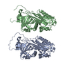
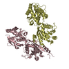
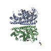
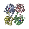
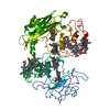
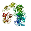
ジャーナル: J Biol Chem / 年: 2019 タイトル: Cryo-EM structure of the native rhodopsin dimer in nanodiscs. 著者: Dorothy Yanling Zhao / Matthias Pöge / Takefumi Morizumi / Sahil Gulati / Ned Van Eps / Jianye Zhang / Przemyslaw Miszta / Slawomir Filipek / Julia Mahamid / Jürgen M Plitzko / Wolfgang ...著者: Dorothy Yanling Zhao / Matthias Pöge / Takefumi Morizumi / Sahil Gulati / Ned Van Eps / Jianye Zhang / Przemyslaw Miszta / Slawomir Filipek / Julia Mahamid / Jürgen M Plitzko / Wolfgang Baumeister / Oliver P Ernst / Krzysztof Palczewski / 要旨: Imaging of rod photoreceptor outer-segment disc membranes by atomic force microscopy and cryo-electron tomography has revealed that the visual pigment rhodopsin, a prototypical class A G protein- ...Imaging of rod photoreceptor outer-segment disc membranes by atomic force microscopy and cryo-electron tomography has revealed that the visual pigment rhodopsin, a prototypical class A G protein-coupled receptor (GPCR), can organize as rows of dimers. GPCR dimerization and oligomerization offer possibilities for allosteric regulation of GPCR activity, but the detailed structures and mechanism remain elusive. In this investigation, we made use of the high rhodopsin density in the native disc membranes and of a bifunctional cross-linker that preserves the native rhodopsin arrangement by covalently tethering rhodopsins via Lys residue side chains. We purified cross-linked rhodopsin dimers and reconstituted them into nanodiscs for cryo-EM analysis. We present cryo-EM structures of the cross-linked rhodopsin dimer as well as a rhodopsin dimer reconstituted into nanodiscs from purified monomers. We demonstrate the presence of a preferential 2-fold symmetrical dimerization interface mediated by transmembrane helix 1 and the cytoplasmic helix 8 of rhodopsin. We confirmed this dimer interface by double electron-electron resonance measurements of spin-labeled rhodopsin. We propose that this interface and the arrangement of two protomers is a prerequisite for the formation of the observed rows of dimers. We anticipate that the approach outlined here could be extended to other GPCRs or membrane receptors to better understand specific receptor dimerization mechanisms.
履歴
登録
2019年3月30日
登録サイト: RCSB / 処理サイト: RCSB
改定 1.0
2019年8月21日
Provider: repository / タイプ: Initial release
改定 1.0
2019年8月21日
Data content type: Additional map / Data content type: Additional map / Provider: repository / タイプ: Initial release
改定 1.0
2019年8月21日
Data content type: EM metadata / Data content type: EM metadata / Provider: repository / タイプ: Initial release
改定 1.0
2019年8月21日
Data content type: Half map / Part number: 1 / Data content type: Half map / Provider: repository / タイプ: Initial release
改定 1.0
2019年8月21日
Data content type: Half map / Part number: 2 / Data content type: Half map / Provider: repository / タイプ: Initial release
改定 1.0
2019年8月21日
Data content type: Image / Data content type: Image / Provider: repository / タイプ: Initial release
改定 1.0
2019年8月21日
Data content type: Mask / Data content type: Mask / Provider: repository / タイプ: Initial release
改定 1.0
2019年8月21日
Data content type: Primary map / Data content type: Primary map / Provider: repository / タイプ: Initial release
改定 1.0
2019年8月21日
Data content type: Additional map / Data content type: Additional map / Provider: repository / タイプ: Initial release
改定 1.0
2019年8月21日
Data content type: EM metadata / Data content type: EM metadata / Provider: repository / タイプ: Initial release
改定 1.0
2019年8月21日
Data content type: Half map / Part number: 1 / Data content type: Half map / Provider: repository / タイプ: Initial release
改定 1.0
2019年8月21日
Data content type: Half map / Part number: 2 / Data content type: Half map / Provider: repository / タイプ: Initial release
改定 1.0
2019年8月21日
Data content type: Image / Data content type: Image / Provider: repository / タイプ: Initial release
改定 1.0
2019年8月21日
Data content type: Mask / Data content type: Mask / Provider: repository / タイプ: Initial release
改定 1.0
2019年8月21日
Data content type: Primary map / Data content type: Primary map / Provider: repository / タイプ: Initial release
Data content type: EM metadata / Data content type: EM metadata / EM metadata / Group: Data processing / Experimental summary / Data content type: EM metadata / EM metadata / カテゴリ: em_admin / em_software / Data content type: EM metadata / EM metadata / Item: _em_admin.last_update / _em_software.name
 ムービー
ムービー コントローラー


 データを開く
 基本情報
基本情報 要素
要素 キーワード
キーワード 機能・相同性情報
 データ登録者
 米国, 1件
  引用
引用


 構造の表示
構造の表示 ダウンロードとリンク
 その他のダウンロード
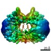


 PDBj
PDBj


 集合体
集合体
 試料調製
試料調製 電子顕微鏡撮影
 FIELD EMISSION GUN / 加速電圧: 300 kV / 照射モード: OTHER
 解析
解析