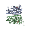
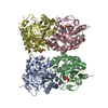
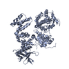
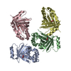
- PDB-3eqx: CRYSTAL STRUCTURE OF A FIC FAMILY PROTEIN (SO_4266) FROM SHEWANEL... -
+
Open data
ID or keywords:
Loading...
-
Basic information
Entry
Database: PDB / ID: 3eqx
Title
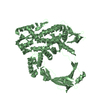
CRYSTAL STRUCTURE OF A FIC FAMILY PROTEIN (SO_4266) FROM SHEWANELLA ONEIDENSIS AT 1.6 A RESOLUTION
Components
FIC DOMAIN CONTAINING TRANSCRIPTIONAL REGULATOR
Keywords
DNA BINDING PROTEIN / FIC FAMILY PROTEIN / STRUCTURAL GENOMICS / JOINT CENTER FOR STRUCTURAL GENOMICS / JCSG / PROTEIN STRUCTURE INITIATIVE / PSI-2
Function / homology
Function and homology information
protein adenylylation / AMPylase activity / protein adenylyltransferase / magnesium ion binding / protein homodimerization activity / ATP binding Similarity search - Function
Fic/DOC N-terminal / Protein adenylyltransferase SoFic-like / : / Fic/DOC family N-terminal / Protein adenylyltransferase SoFic-like, C-terminal domain / Fido domain-containing protein / Fido-like domain superfamily / Fic/DOC family / Fido domain / Fido domain profile. Similarity search - Domain/homology
Mass: 18.015 Da / Num. of mol.: 814 / Source method: isolated from a natural source / Formula: H2O
Has protein modification
Y
Sequence details
THE CONSTRUCT WAS EXPRESSED WITH A PURIFICATION TAG MGSDKIHHHHHHENLYFQG. THE TAG WAS REMOVED WITH ...THE CONSTRUCT WAS EXPRESSED WITH A PURIFICATION TAG MGSDKIHHHHHHENLYFQG. THE TAG WAS REMOVED WITH TEV PROTEASE LEAVING ONLY A GLYCINE (0) FOLLOWED BY THE TARGET SEQUENCE. DNA SEQUENCING OF THE CLONED CONSTRUCT SHOWS A CYSTEINE AT POSITION 109 INSTEAD OF A GLYCINE. THE CYSTEINE AT POSITION 109 IS SUPPORTED BY THE ELECTRON DENSITY.
-
Experimental details
-
Experiment
Experiment
Method: X-RAY DIFFRACTION / Number of used crystals: 1
-
Sample preparation
Crystal
Density Matthews: 2.36 Å3/Da / Density % sol: 47.95 %
Crystal grow
Temperature: 277 K / Method: vapor diffusion, sitting drop / pH: 7.1 Details: 0.2M NaF, 20.0% PEG-3350, No Buffer pH 7.1, NANODROP, VAPOR DIFFUSION, SITTING DROP, temperature 277K
Type: MARMOSAIC 325 mm CCD / Detector: CCD / Date: May 13, 2007 / Details: Flat mirror (vertical focusing)
Radiation
Monochromator: Single crystal Si(111) bent monochromator (horizontal focusing) Protocol: MAD / Monochromatic (M) / Laue (L): M / Scattering type: x-ray
Radiation wavelength
ID
Wavelength (Å)
Relative weight
1
0.91837
1
2
0.97905
1
3
0.97935
1
Reflection
Resolution: 1.6→29.553 Å / Num. obs: 106471 / % possible obs: 98.7 % / Observed criterion σ(I): -3 / Biso Wilson estimate: 19.465 Å2 / Rmerge(I) obs: 0.042 / Net I/σ(I): 13.83
Reflection shell
Resolution (Å)
Rmerge(I) obs
Mean I/σ(I) obs
Num. measured obs
Num. unique obs
Diffraction-ID
% possible all
1.6-1.66
0.524
2
37525
21263
1
98.7
1.66-1.72
0.415
2.4
33144
18630
1
99.7
1.72-1.8
0.327
3.1
37611
21027
1
99.6
1.8-1.9
0.234
4.3
38912
21626
1
99.7
1.9-2.02
0.158
6.3
37195
20547
1
99.5
2.02-2.17
0.102
9.4
35933
19785
1
99.7
2.17-2.39
0.066
13.9
37908
20659
1
99.3
2.39-2.73
0.043
20
37476
20049
1
98.7
2.73-3.44
0.026
29.6
38826
20295
1
97.7
3.44-29.553
0.015
48.8
38572
19736
1
94.9
-
Phasing
Phasing
Method: MAD
-
Processing
Software
Name
Version
Classification
NB
REFMAC
5.2.0019
refinement
PHENIX
refinement
SHELX
phasing
MolProbity
3beta29
modelbuilding
XSCALE
datascaling
PDB_EXTRACT
3.006
dataextraction
XDS
datareduction
SOLVE
phasing
Refinement
Method to determine structure: MAD / Resolution: 1.6→29.553 Å / Cor.coef. Fo:Fc: 0.966 / Cor.coef. Fo:Fc free: 0.955 / Occupancy max: 1 / Occupancy min: 0.25 / SU B: 3.098 / SU ML: 0.055 / TLS residual ADP flag: LIKELY RESIDUAL / Cross valid method: THROUGHOUT / σ(F): 0 / ESU R: 0.084 / ESU R Free: 0.083 Stereochemistry target values: MAXIMUM LIKELIHOOD WITH PHASES Details: 1. HYDROGENS HAVE BEEN ADDED IN THE RIDING POSITIONS 2. A MET-INHIBITION PROTOCOL WAS USED FOR SELENOMETHIONINE INCORPORATION DURING PROTEIN EXPRESSION. THE OCCUPANCY OF THE SE ATOMS IN THE ...Details: 1. HYDROGENS HAVE BEEN ADDED IN THE RIDING POSITIONS 2. A MET-INHIBITION PROTOCOL WAS USED FOR SELENOMETHIONINE INCORPORATION DURING PROTEIN EXPRESSION. THE OCCUPANCY OF THE SE ATOMS IN THE MSE RESIDUES WAS REDUCED TO 0.75 TO ACCOUNT FOR THE REDUCED SCATTERING POWER DUE TO PARTIAL S-MET INCORPORATION 3. ATOM RECORD CONTAINS RESIDUAL B FACTORS ONLY. 4. MODELING EXPERIMENTS SUGGEST THAT THE ELECTRON DENSITY NEAR THE HPFXXGNG MOTIF (PRESENT IN FIC PROTEINS) STARTING AT HIS198 IN CHAIN A IS LIKELY TO BE A PORTION (B1-B4) OF THE PARTIALLY MISSING N-TERMINUS OF CHAIN B FROM A SYMMETRY RELATED MOLECULE. B1(MSE) IS SUPPORTED BY A SIGNIGICANT PEAK IN THE ANOMALOUS DIFFERENCE FOURIER MAP CALCULATED FOR THIS SELENOMETHIONINE DERIVATIZED PROTEIN CRYSTAL. 5. PGE HAS BEEN MODELED BASED ON CRYSTALLIZATION CONDITIONS. 6. RESIDUES A344-A345 IN CHAIN A AND B5-B10 ARE DISORDERED AND HAVE NOT BEEN MODELLED.
Rfactor
Num. reflection
% reflection
Selection details
Rfree
0.197
5304
5 %
RANDOM
Rwork
0.168
-
-
-
obs
0.17
106418
99.32 %
-
Solvent computation
Ion probe radii: 0.8 Å / Shrinkage radii: 0.8 Å / VDW probe radii: 1.2 Å / Solvent model: MASK
In the structure databanks used in Yorodumi, some data are registered as the other names, "COVID-19 virus" and "2019-nCoV". Here are the details of the virus and the list of structure data.
Jan 31, 2019. EMDB accession codes are about to change! (news from PDBe EMDB page)
EMDB accession codes are about to change! (news from PDBe EMDB page)
The allocation of 4 digits for EMDB accession codes will soon come to an end. Whilst these codes will remain in use, new EMDB accession codes will include an additional digit and will expand incrementally as the available range of codes is exhausted. The current 4-digit format prefixed with “EMD-” (i.e. EMD-XXXX) will advance to a 5-digit format (i.e. EMD-XXXXX), and so on. It is currently estimated that the 4-digit codes will be depleted around Spring 2019, at which point the 5-digit format will come into force.
The EM Navigator/Yorodumi systems omit the EMD- prefix.
Related info.:Q: What is EMD? / ID/Accession-code notation in Yorodumi/EM Navigator
Yorodumi is a browser for structure data from EMDB, PDB, SASBDB, etc.
This page is also the successor to EM Navigator detail page, and also detail information page/front-end page for Omokage search.
The word "yorodu" (or yorozu) is an old Japanese word meaning "ten thousand". "mi" (miru) is to see.
Related info.:EMDB / PDB / SASBDB / Comparison of 3 databanks / Yorodumi Search / Aug 31, 2016. New EM Navigator & Yorodumi / Yorodumi Papers / Jmol/JSmol / Function and homology information / Changes in new EM Navigator and Yorodumi
 Movie
Movie Controller

 Yorodumi
 Open data
 Basic information
 Components
 Keywords
 Function and homology information
 Shewanella oneidensis (bacteria)
 X-RAY DIFFRACTION /
  Authors
 Citation
 Structure visualization
 Downloads & links
 Other downloads


 PDBj
PDBj Assembly
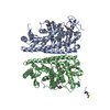

 Mass: 150.173 Da / Num. of mol.: 1 / Source method: obtained synthetically / Formula: C6H14O4
 Mass: 18.015 Da / Num. of mol.: 814 / Source method: isolated from a natural source / Formula: H2O
 Sample preparation
 / Beamline: BL11-1 / Wavelength: 0.91837,0.97905,0.97935
 Processing