 Movie
Movie Controller


+ Open data

- Basic information
| Entry | Database: PDB / ID: 4dvz | ||||||
|---|---|---|---|---|---|---|---|
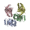
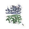
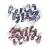
| Title | Crystal structure of the Helicobacter pylori CagA oncoprotein | ||||||
 Components Components | Cytotoxicity-associated immunodominant antigen | ||||||
 Keywords Keywords | ONCOPROTEIN | ||||||
| Function / homology |  Function and homology information Function and homology informationsymbiont-mediated perturbation of host cell cycle progression / toxin transmembrane transporter activity / molecular adaptor activity Similarity search - Function | ||||||
| Biological species |   Helicobacter pylori (bacteria) Helicobacter pylori (bacteria) | ||||||
| Method |  X-RAY DIFFRACTION / SYNCHROTRON / MAD / Resolution: 3.19 Å X-RAY DIFFRACTION / SYNCHROTRON / MAD / Resolution: 3.19 Å | ||||||
 Authors Authors | Hayashi, T. / Senda, M. / Morohashi, H. / Higashi, H. / Horio, M. / Kashiba, Y. / Nagase, L. / Sasaya, D. / Shimizu, T. / Venugopalan, N. ...Hayashi, T. / Senda, M. / Morohashi, H. / Higashi, H. / Horio, M. / Kashiba, Y. / Nagase, L. / Sasaya, D. / Shimizu, T. / Venugopalan, N. / Kumeta, H. / Noda, N. / Inagaki, F. / Senda, T. / Hatakeyama, M. | ||||||
 Citation Citation | Journal: Cell Host Microbe / Year: 2012 Title: Tertiary structure-function analysis reveals the pathogenic signaling potentiation mechanism of Helicobacter pylori oncogenic effector CagA Authors: Hayashi, T. / Senda, M. / Morohashi, H. / Higashi, H. / Horio, M. / Kashiba, Y. / Nagase, L. / Sasaya, D. / Shimizu, T. / Venugopalan, N. / Kumeta, H. / Noda, N.N. / Inagaki, F. / Senda, T. / Hatakeyama, M. | ||||||
| History |
|
- Structure visualization
| Structure viewer | Molecule: MolmilJmol/JSmol |
|---|
- Downloads & links
-Download
| PDBx/mmCIF format | 4dvz.cif.gz | 103.3 KB | Display | PDBx/mmCIF format |
|---|---|---|---|---|
| PDB format | pdb4dvz.ent.gz | 78.5 KB | Display | PDB format |
| PDBx/mmJSON format | 4dvz.json.gz | Tree view | PDBx/mmJSON format | |
| Others |  Other downloads Other downloads |
-Validation report
| Arichive directory | https://data.pdbj.org/pub/pdb/validation_reports/dv/4dvzftp://data.pdbj.org/pub/pdb/validation_reports/dv/4dvz | HTTPS FTP |
|---|
-Related structure data


-Links
 PDBj
PDBj- Assembly
| Deposited unit | 
| ||||||||
|---|---|---|---|---|---|---|---|---|---|
| 1 |
| ||||||||
| Unit cell |
|
-Components
| #1: Protein | Mass: 63625.383 Da / Num. of mol.: 1 / Fragment: UNP residues 261-829 Source method: isolated from a genetically manipulated source Source: (gene. exp.) Helicobacter pylori (bacteria) / Strain: 26695 / Gene: cagA / Plasmid: pGEX-6P-2 / Production host: |
|---|
-Experimental details
-Experiment
| Experiment | Method: X-RAY DIFFRACTION / Number of used crystals: 1 |
|---|
- Sample preparation
| Crystal | Density Matthews: 3.49 Å3/Da / Density % sol: 64.72 % |
|---|---|
| Crystal grow | Temperature: 293 K / Method: vapor diffusion, sitting drop / pH: 7 Details: 7% EtOH, 50mM Tris-HCl, pH 7.0, VAPOR DIFFUSION, SITTING DROP, temperature 293K |
-Data collection
| Diffraction | Mean temperature: 95 K | |||||||||
|---|---|---|---|---|---|---|---|---|---|---|
| Diffraction source | Source: SYNCHROTRON / Site: Photon Factory  / Beamline: AR-NE3A / Wavelength: 0.98057, 0.98086 / Beamline: AR-NE3A / Wavelength: 0.98057, 0.98086 | |||||||||
| Detector | Type: ADSC QUANTUM 270 / Detector: CCD / Date: Mar 6, 2011 | |||||||||
| Radiation | Monochromator: Si 111 / Protocol: MAD / Monochromatic (M) / Laue (L): M / Scattering type: x-ray | |||||||||
| Radiation wavelength |
| |||||||||
| Reflection | Resolution: 3.19→74.3 Å / Num. all: 15242 / Num. obs: 15242 / % possible obs: 99.3 % / Observed criterion σ(F): 0 / Observed criterion σ(I): 0 / Redundancy: 10.6 % / Biso Wilson estimate: 97.02 Å2 / Rmerge(I) obs: 0.059 / Net I/σ(I): 33.39 | |||||||||
| Reflection shell | Resolution: 3.19→3.37 Å / Redundancy: 11.1 % / Rmerge(I) obs: 0.468 / Mean I/σ(I) obs: 6 / Num. unique all: 2263 / % possible all: 100 |
- Processing
| Software |
| ||||||||||||||||||||||||||||||||||||||||||||||||||||||||||||||||||||||||||||||||||||||||||||||||||||||||||||
|---|---|---|---|---|---|---|---|---|---|---|---|---|---|---|---|---|---|---|---|---|---|---|---|---|---|---|---|---|---|---|---|---|---|---|---|---|---|---|---|---|---|---|---|---|---|---|---|---|---|---|---|---|---|---|---|---|---|---|---|---|---|---|---|---|---|---|---|---|---|---|---|---|---|---|---|---|---|---|---|---|---|---|---|---|---|---|---|---|---|---|---|---|---|---|---|---|---|---|---|---|---|---|---|---|---|---|---|---|---|
| Refinement | Method to determine structure: MAD / Resolution: 3.19→74.3 Å / Cor.coef. Fo:Fc: 0.9219 / Cor.coef. Fo:Fc free: 0.8671 / Occupancy max: 1 / Occupancy min: 1 / SU R Cruickshank DPI: 1.611 / Cross valid method: THROUGHOUT / σ(F): 0 / SU R Blow DPI: 1.553 / SU Rfree Blow DPI: 0.426 / SU Rfree Cruickshank DPI: 0.435 / Stereochemistry target values: BUSTER
| ||||||||||||||||||||||||||||||||||||||||||||||||||||||||||||||||||||||||||||||||||||||||||||||||||||||||||||
| Displacement parameters | Biso max: 186.4 Å2 / Biso mean: 103.74 Å2 / Biso min: 56 Å2
| ||||||||||||||||||||||||||||||||||||||||||||||||||||||||||||||||||||||||||||||||||||||||||||||||||||||||||||
| Refine analyze | Luzzati coordinate error obs: 0.799 Å | ||||||||||||||||||||||||||||||||||||||||||||||||||||||||||||||||||||||||||||||||||||||||||||||||||||||||||||
| Refinement step | Cycle: LAST / Resolution: 3.19→74.3 Å
| ||||||||||||||||||||||||||||||||||||||||||||||||||||||||||||||||||||||||||||||||||||||||||||||||||||||||||||
| Refine LS restraints |
| ||||||||||||||||||||||||||||||||||||||||||||||||||||||||||||||||||||||||||||||||||||||||||||||||||||||||||||
| LS refinement shell | Resolution: 3.19→3.41 Å / Total num. of bins used: 8
|
