Movie
Movie Controller


+ Open data

- Basic information
| Entry | Database: PDB / ID: 6o96 | ||||||||||||
|---|---|---|---|---|---|---|---|---|---|---|---|---|---|
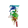
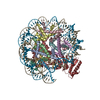
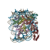
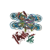
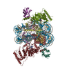
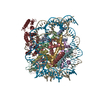
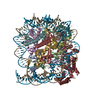
| Title | Dot1L bound to the H2BK120 Ubiquitinated nucleosome | ||||||||||||
 Components Components |
| ||||||||||||
 Keywords Keywords | Structural Protein/DNA/Transferase / Complex / Chromatin Modifier / Structural Protein / TRANSFERASE / Structural Protein-DNA-Transferase complex | ||||||||||||
| Function / homology |  Function and homology information Function and homology information[histone H3]-lysine79 N-trimethyltransferase / histone H3K79 methyltransferase activity / histone H3K79 trimethyltransferase activity / regulation of transcription regulatory region DNA binding / hypothalamus gonadotrophin-releasing hormone neuron development / female meiosis I / positive regulation of protein monoubiquitination / regulation of receptor signaling pathway via JAK-STAT / fat pad development / mitochondrion transport along microtubule ...[histone H3]-lysine79 N-trimethyltransferase / histone H3K79 methyltransferase activity / histone H3K79 trimethyltransferase activity / regulation of transcription regulatory region DNA binding / hypothalamus gonadotrophin-releasing hormone neuron development / female meiosis I / positive regulation of protein monoubiquitination / regulation of receptor signaling pathway via JAK-STAT / fat pad development / mitochondrion transport along microtubule / histone H3 methyltransferase activity / histone methyltransferase activity / female gonad development / seminiferous tubule development / male meiosis I / positive regulation of intrinsic apoptotic signaling pathway by p53 class mediator / subtelomeric heterochromatin formation / energy homeostasis / regulation of neuron apoptotic process / neuron projection morphogenesis / regulation of proteasomal protein catabolic process / Maturation of protein E / Maturation of protein E / ER Quality Control Compartment (ERQC) / Myoclonic epilepsy of Lafora / FLT3 signaling by CBL mutants / IRAK2 mediated activation of TAK1 complex / Prevention of phagosomal-lysosomal fusion / Alpha-protein kinase 1 signaling pathway / Glycogen synthesis / IRAK1 recruits IKK complex / IRAK1 recruits IKK complex upon TLR7/8 or 9 stimulation / Endosomal Sorting Complex Required For Transport (ESCRT) / Membrane binding and targetting of GAG proteins / Negative regulation of FLT3 / Regulation of TBK1, IKKε (IKBKE)-mediated activation of IRF3, IRF7 / PTK6 Regulates RTKs and Their Effectors AKT1 and DOK1 / Regulation of TBK1, IKKε-mediated activation of IRF3, IRF7 upon TLR3 ligation / Constitutive Signaling by NOTCH1 HD Domain Mutants / IRAK2 mediated activation of TAK1 complex upon TLR7/8 or 9 stimulation / NOTCH2 Activation and Transmission of Signal to the Nucleus / TICAM1,TRAF6-dependent induction of TAK1 complex / TICAM1-dependent activation of IRF3/IRF7 / APC/C:Cdc20 mediated degradation of Cyclin B / telomere organization / Downregulation of ERBB4 signaling / Regulation of FZD by ubiquitination / APC-Cdc20 mediated degradation of Nek2A / p75NTR recruits signalling complexes / InlA-mediated entry of Listeria monocytogenes into host cells / TRAF6 mediated IRF7 activation in TLR7/8 or 9 signaling / TRAF6-mediated induction of TAK1 complex within TLR4 complex / Regulation of pyruvate metabolism / NF-kB is activated and signals survival / Regulation of innate immune responses to cytosolic DNA / Pexophagy / Downregulation of ERBB2:ERBB3 signaling / NRIF signals cell death from the nucleus / VLDLR internalisation and degradation / Regulation of PTEN localization / DNA damage checkpoint signaling / Activated NOTCH1 Transmits Signal to the Nucleus / Regulation of BACH1 activity / Synthesis of active ubiquitin: roles of E1 and E2 enzymes / MAP3K8 (TPL2)-dependent MAPK1/3 activation / Translesion synthesis by REV1 / TICAM1, RIP1-mediated IKK complex recruitment / Translesion synthesis by POLK / InlB-mediated entry of Listeria monocytogenes into host cell / Activation of IRF3, IRF7 mediated by TBK1, IKKε (IKBKE) / JNK (c-Jun kinases) phosphorylation and activation mediated by activated human TAK1 / Downregulation of TGF-beta receptor signaling / Josephin domain DUBs / positive regulation of protein ubiquitination / Translesion synthesis by POLI / Gap-filling DNA repair synthesis and ligation in GG-NER / IKK complex recruitment mediated by RIP1 / Regulation of activated PAK-2p34 by proteasome mediated degradation / PINK1-PRKN Mediated Mitophagy / TGF-beta receptor signaling in EMT (epithelial to mesenchymal transition) / TNFR1-induced NF-kappa-B signaling pathway / regulation of mitochondrial membrane potential / Autodegradation of Cdh1 by Cdh1:APC/C / TCF dependent signaling in response to WNT / APC/C:Cdc20 mediated degradation of Securin / Regulation of NF-kappa B signaling / N-glycan trimming in the ER and Calnexin/Calreticulin cycle / Asymmetric localization of PCP proteins / activated TAK1 mediates p38 MAPK activation / Ubiquitin-dependent degradation of Cyclin D / SCF-beta-TrCP mediated degradation of Emi1 / NIK-->noncanonical NF-kB signaling / TNFR2 non-canonical NF-kB pathway / AUF1 (hnRNP D0) binds and destabilizes mRNA / Regulation of signaling by CBL / NOTCH3 Activation and Transmission of Signal to the Nucleus / Negative regulators of DDX58/IFIH1 signaling / Vpu mediated degradation of CD4 / Assembly of the pre-replicative complex / Ubiquitin-Mediated Degradation of Phosphorylated Cdc25A Similarity search - Function | ||||||||||||
| Biological species | synthetic construct (others)  Homo sapiens (human) Homo sapiens (human) | ||||||||||||
| Method | ELECTRON MICROSCOPY / single particle reconstruction / cryo EM / Resolution: 3.5 Å | ||||||||||||
 Authors Authors | Valencia-Sanchez, M.I. / De Ioannes, P.E. / Miao, W. / Vasilyev, N. / Chen, R. / Nudler, E. / Armache, J.-P. / Armache, K.-J. | ||||||||||||
| Funding support |  United States, 3items United States, 3items
| ||||||||||||
 Citation Citation | Journal: Mol Cell / Year: 2019 Title: Structural Basis of Dot1L Stimulation by Histone H2B Lysine 120 Ubiquitination. Authors: Marco Igor Valencia-Sánchez / Pablo De Ioannes / Miao Wang / Nikita Vasilyev / Ruoyu Chen / Evgeny Nudler / Jean-Paul Armache / Karim-Jean Armache / Abstract: The essential histone H3 lysine 79 methyltransferase Dot1L regulates transcription and genomic stability and is deregulated in leukemia. The activity of Dot1L is stimulated by mono-ubiquitination of ...The essential histone H3 lysine 79 methyltransferase Dot1L regulates transcription and genomic stability and is deregulated in leukemia. The activity of Dot1L is stimulated by mono-ubiquitination of histone H2B on lysine 120 (H2BK120Ub); however, the detailed mechanism is not understood. We report cryo-EM structures of human Dot1L bound to (1) H2BK120Ub and (2) unmodified nucleosome substrates at 3.5 Å and 4.9 Å, respectively. Comparison of both structures, complemented with biochemical experiments, provides critical insights into the mechanism of Dot1L stimulation by H2BK120Ub. Both structures show Dot1L binding to the same extended surface of the histone octamer. In yeast, this surface is used by silencing proteins involved in heterochromatin formation, explaining the mechanism of their competition with Dot1. These results provide a strong foundation for understanding conserved crosstalk between histone modifications found at actively transcribed genes and offer a general model of how ubiquitin might regulate the activity of chromatin enzymes. | ||||||||||||
| History |
|
- Structure visualization
| Movie |
Movie viewer |
|---|---|
| Structure viewer | Molecule: MolmilJmol/JSmol |
- Downloads & links
-Download
| PDBx/mmCIF format | 6o96.cif.gz | 361.4 KB | Display | PDBx/mmCIF format |
|---|---|---|---|---|
| PDB format | pdb6o96.ent.gz | 270.8 KB | Display | PDB format |
| PDBx/mmJSON format | 6o96.json.gz | Tree view | PDBx/mmJSON format | |
| Others |  Other downloads Other downloads |
-Validation report
| Summary document | 6o96_validation.pdf.gz | 1.1 MB | Display | wwPDB validaton report |
|---|---|---|---|---|
| Full document | 6o96_full_validation.pdf.gz | 1.1 MB | Display | |
| Data in XML | 6o96_validation.xml.gz | 47 KB | Display | |
| Data in CIF | 6o96_validation.cif.gz | 72.5 KB | Display | |
| Arichive directory | https://data.pdbj.org/pub/pdb/validation_reports/o9/6o96ftp://data.pdbj.org/pub/pdb/validation_reports/o9/6o96 | HTTPS FTP |
-Related structure data
| Related structure data | 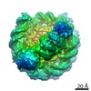 0652MC 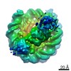 0653C 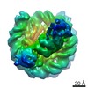 0654C  0655C M: map data used to model this data C: citing same article ( |
|---|---|
| Similar structure data |
-Links
 PDBj
PDBj


- Assembly
| Deposited unit | 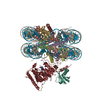
|
|---|---|
| 1 |
|
-Components
-Protein , 6 types, 10 molecules AEBFCGDHKL
| #1: Protein | Mass: 15303.930 Da / Num. of mol.: 2 / Mutation: G102A Source method: isolated from a genetically manipulated source Source: (gene. exp.)  #2: Protein | Mass: 11394.426 Da / Num. of mol.: 2 Source method: isolated from a genetically manipulated source Source: (gene. exp.) #3: Protein | Mass: 14109.436 Da / Num. of mol.: 2 / Mutation: G99R, A123S Source method: isolated from a genetically manipulated source Source: (gene. exp.) #4: Protein | Mass: 13629.911 Da / Num. of mol.: 2 / Mutation: K120C, S32T Source method: isolated from a genetically manipulated source Source: (gene. exp.) #7: Protein | | Mass: 38245.379 Da / Num. of mol.: 1 Source method: isolated from a genetically manipulated source Source: (gene. exp.) Homo sapiens (human) / Gene: DOT1L, KIAA1814, KMT4 / Production host: References: UniProt: Q8TEK3, histone-lysine N-methyltransferase #8: Protein | | Mass: 8622.922 Da / Num. of mol.: 1 / Mutation: G76C Source method: isolated from a genetically manipulated source Source: (gene. exp.) Homo sapiens (human) / Gene: UBB / Production host: |
|---|
-DNA chain , 2 types, 2 molecules IJ
| #5: DNA chain | Mass: 44824.570 Da / Num. of mol.: 1 Source method: isolated from a genetically manipulated source Source: (gene. exp.) synthetic construct (others) / Production host: |
|---|---|
| #6: DNA chain | Mass: 45304.863 Da / Num. of mol.: 1 Source method: isolated from a genetically manipulated source Source: (gene. exp.) synthetic construct (others) / Production host: |
-Non-polymers , 1 types, 1 molecules 
| #9: Chemical | ChemComp-SAH /  Mass: 384.411 Da / Num. of mol.: 1 / Source method: obtained synthetically / Formula: C14H20N6O5S Mass: 384.411 Da / Num. of mol.: 1 / Source method: obtained synthetically / Formula: C14H20N6O5S |
|---|
-Details
| Has protein modification | N |
|---|
-Experimental details
-Experiment
| Experiment | Method: ELECTRON MICROSCOPY |
|---|---|
| EM experiment | Aggregation state: PARTICLE / 3D reconstruction method: single particle reconstruction |
- Sample preparation
| Component |
| |||||||||||||||||||||||||||||||||||||||||||||||||
|---|---|---|---|---|---|---|---|---|---|---|---|---|---|---|---|---|---|---|---|---|---|---|---|---|---|---|---|---|---|---|---|---|---|---|---|---|---|---|---|---|---|---|---|---|---|---|---|---|---|---|
| Molecular weight |
| |||||||||||||||||||||||||||||||||||||||||||||||||
| Source (natural) |
| |||||||||||||||||||||||||||||||||||||||||||||||||
| Source (recombinant) |
| |||||||||||||||||||||||||||||||||||||||||||||||||
| Buffer solution | pH: 7 | |||||||||||||||||||||||||||||||||||||||||||||||||
| Specimen | Conc.: 0.5 mg/ml / Embedding applied: NO / Shadowing applied: NO / Staining applied: NO / Vitrification applied: YES / Details: 3.5 angstroms Dot1L-H2BK120Ub Sample 1A | |||||||||||||||||||||||||||||||||||||||||||||||||
| Specimen support | Grid material: GOLD / Grid mesh size: 200 divisions/in. / Grid type: Quantifoil R1.2/1.3 | |||||||||||||||||||||||||||||||||||||||||||||||||
| Vitrification | Instrument: FEI VITROBOT MARK IV / Cryogen name: PROPANE / Humidity: 100 % / Chamber temperature: 297.15 K / Details: blotted for 3s before plunging |
- Electron microscopy imaging
| Experimental equipment |  Model: Titan Krios / Image courtesy: FEI Company |
|---|---|
| Microscopy | Model: FEI TITAN KRIOS |
| Electron gun | Electron source:  FIELD EMISSION GUN / Accelerating voltage: 300 kV / Illumination mode: FLOOD BEAM FIELD EMISSION GUN / Accelerating voltage: 300 kV / Illumination mode: FLOOD BEAM |
| Electron lens | Mode: BRIGHT FIELD / Nominal magnification: 130000 X / Nominal defocus max: 3200 nm / Nominal defocus min: 1700 nm / Cs: 2.7 mm |
| Specimen holder | Cryogen: NITROGEN |
| Image recording | Electron dose: 1.1 e/Å2 / Detector mode: SUPER-RESOLUTION / Film or detector model: GATAN K2 SUMMIT (4k x 4k) |
- Processing
| Software | Name: PHENIX / Version: 1.14_3260: / Classification: refinement | ||||||||||||||||||||||||||||
|---|---|---|---|---|---|---|---|---|---|---|---|---|---|---|---|---|---|---|---|---|---|---|---|---|---|---|---|---|---|
| EM software |
| ||||||||||||||||||||||||||||
| CTF correction | Type: PHASE FLIPPING AND AMPLITUDE CORRECTION | ||||||||||||||||||||||||||||
| Symmetry | Point symmetry: C1 (asymmetric) | ||||||||||||||||||||||||||||
| 3D reconstruction | Resolution: 3.5 Å / Resolution method: FSC 0.143 CUT-OFF / Num. of particles: 211279 / Symmetry type: POINT | ||||||||||||||||||||||||||||
| Atomic model building | B value: 91.61 / Protocol: OTHER / Space: REAL | ||||||||||||||||||||||||||||
| Atomic model building | 3D fitting-ID: 1 / Pdb chain-ID: A / Source name: PDB / Type: experimental model
| ||||||||||||||||||||||||||||
| Refine LS restraints |
|