Movie
Movie Controller


+ Open data

- Basic information
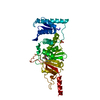
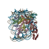
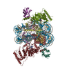
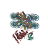
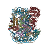
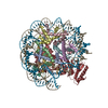
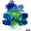
| Entry | Database: PDB / ID: 6nn6 | |||||||||
|---|---|---|---|---|---|---|---|---|---|---|
| Title | Structure of Dot1L-H2BK120ub nucleosome complex | |||||||||
 Components Components |
| |||||||||
 Keywords Keywords | GENE REGULATION / chromatin / structural biology / single particle / cryo-EM / histone methyltransferase / nucleosome / Dot1L | |||||||||
| Function / homology |  Function and homology information Function and homology information[histone H3]-lysine79 N-trimethyltransferase / histone H3K79 trimethyltransferase activity / histone H3K79 methyltransferase activity / regulation of transcription regulatory region DNA binding / regulation of receptor signaling pathway via JAK-STAT / histone H3 methyltransferase activity / histone methyltransferase activity / sodium ion transport / subtelomeric heterochromatin formation / DNA damage checkpoint signaling ...[histone H3]-lysine79 N-trimethyltransferase / histone H3K79 trimethyltransferase activity / histone H3K79 methyltransferase activity / regulation of transcription regulatory region DNA binding / regulation of receptor signaling pathway via JAK-STAT / histone H3 methyltransferase activity / histone methyltransferase activity / sodium ion transport / subtelomeric heterochromatin formation / DNA damage checkpoint signaling / telomere organization / PKMTs methylate histone lysines / structural constituent of chromatin / nucleosome / nucleosome assembly / heterochromatin formation / gene expression / methylation / nucleic acid binding / RNA polymerase II-specific DNA-binding transcription factor binding / transcription coactivator activity / mitochondrial outer membrane / chromosome, telomeric region / protein heterodimerization activity / DNA repair / positive regulation of transcription by RNA polymerase II / protein-containing complex / DNA binding / nucleoplasm / nucleus / cytoplasm Similarity search - Function | |||||||||
| Biological species |  Homo sapiens (human) Homo sapiens (human)synthetic construct (others) | |||||||||
| Method | ELECTRON MICROSCOPY / single particle reconstruction / cryo EM / Resolution: 3.9 Å | |||||||||
 Authors Authors | Anderson, C.J. / Baird, M.R. / Hsu, A. / Barbour, E.H. / Koyama, Y. / Borgnia, M.J. / McGinty, R.K. | |||||||||
| Funding support |  United States, 2items United States, 2items
| |||||||||
 Citation Citation | Journal: Cell Rep / Year: 2019 Title: Structural Basis for Recognition of Ubiquitylated Nucleosome by Dot1L Methyltransferase. Authors: Cathy J Anderson / Matthew R Baird / Allen Hsu / Emily H Barbour / Yuka Koyama / Mario J Borgnia / Robert K McGinty / Abstract: Histone H3 lysine 79 (H3K79) methylation is enriched on actively transcribed genes, and its misregulation is a hallmark of leukemia. Methylation of H3K79, which resides on the structured disk face of ...Histone H3 lysine 79 (H3K79) methylation is enriched on actively transcribed genes, and its misregulation is a hallmark of leukemia. Methylation of H3K79, which resides on the structured disk face of the nucleosome, is mediated by the Dot1L methyltransferase. Dot1L activity is part of a trans-histone crosstalk pathway, requiring prior histone H2B ubiquitylation of lysine 120 (H2BK120ub) for optimal activity. However, the molecular details describing both how Dot1L binds to the nucleosome and why Dot1L is activated by H2BK120 ubiquitylation are unknown. Here, we present the cryoelectron microscopy (cryo-EM) structure of Dot1L bound to a nucleosome reconstituted with site-specifically ubiquitylated H2BK120. The structure reveals that Dot1L engages the nucleosome acidic patch using a variant arginine anchor and occupies a conformation poised for methylation. In this conformation, Dot1L and ubiquitin interact directly through complementary hydrophobic surfaces. This study establishes a path to better understand Dot1L function in normal and leukemia cells. | |||||||||
| History |
|
- Structure visualization
| Movie |
Movie viewer |
|---|---|
| Structure viewer | Molecule: MolmilJmol/JSmol |
- Downloads & links
-Download
| PDBx/mmCIF format | 6nn6.cif.gz | 353.4 KB | Display | PDBx/mmCIF format |
|---|---|---|---|---|
| PDB format | pdb6nn6.ent.gz | 269.3 KB | Display | PDB format |
| PDBx/mmJSON format | 6nn6.json.gz | Tree view | PDBx/mmJSON format | |
| Others |  Other downloads Other downloads |
-Validation report
| Arichive directory | https://data.pdbj.org/pub/pdb/validation_reports/nn/6nn6ftp://data.pdbj.org/pub/pdb/validation_reports/nn/6nn6 | HTTPS FTP |
|---|
-Related structure data
| Related structure data |  0458MC 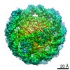 0459C 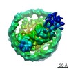 0460C M: map data used to model this data C: citing same article ( |
|---|---|
| Similar structure data |
-Links
 PDBj
PDBj


- Assembly
| Deposited unit | 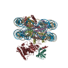
|
|---|---|
| 1 |
|
-Components
-Protein , 6 types, 10 molecules AEBFCGDHKL
| #1: Protein | Mass: 15303.930 Da / Num. of mol.: 2 / Mutation: G103A Source method: isolated from a genetically manipulated source Source: (gene. exp.)  #2: Protein | Mass: 11263.231 Da / Num. of mol.: 2 Source method: isolated from a genetically manipulated source Source: (gene. exp.) #3: Protein | Mass: 13978.241 Da / Num. of mol.: 2 / Mutation: G100R, A124S Source method: isolated from a genetically manipulated source Source: (gene. exp.) #4: Protein | Mass: 13642.846 Da / Num. of mol.: 2 / Mutation: S33T, K121C Source method: isolated from a genetically manipulated source Source: (gene. exp.) #7: Protein | | Mass: 47462.039 Da / Num. of mol.: 1 Source method: isolated from a genetically manipulated source Source: (gene. exp.) Homo sapiens (human) / Gene: DOT1L, KIAA1814, KMT4 / Plasmid: pST50 / Production host: References: UniProt: Q8TEK3, histone-lysine N-methyltransferase #8: Protein | | Mass: 8911.180 Da / Num. of mol.: 1 / Mutation: G76S Source method: isolated from a genetically manipulated source Source: (gene. exp.) Homo sapiens (human) / Gene: UBC / Plasmid: pST50 / Production host: |
|---|
-DNA chain , 2 types, 2 molecules IJ
| #5: DNA chain | Mass: 44520.383 Da / Num. of mol.: 1 / Source method: obtained synthetically / Details: Widom 601 nucleosome positioning sequence / Source: (synth.) synthetic construct (others) |
|---|---|
| #6: DNA chain | Mass: 44991.660 Da / Num. of mol.: 1 / Source method: obtained synthetically / Details: Widom 601 nucleosome positioning sequence / Source: (synth.) synthetic construct (others) |
-Experimental details
-Experiment
| Experiment | Method: ELECTRON MICROSCOPY |
|---|---|
| EM experiment | Aggregation state: PARTICLE / 3D reconstruction method: single particle reconstruction |
- Sample preparation
| Component | Name: Dot1L-H2BK120ub nucleosome complex (Class 2A) / Type: COMPLEX / Entity ID: all / Source: RECOMBINANT |
|---|---|
| Molecular weight | Value: 0.27 MDa / Experimental value: NO |
| Source (natural) | Organism: Homo sapiens (human) |
| Source (recombinant) | Organism: |
| Buffer solution | pH: 7.5 |
| Specimen | Conc.: 0.93 mg/ml / Embedding applied: NO / Shadowing applied: NO / Staining applied: NO / Vitrification applied: YES |
| Specimen support | Grid material: COPPER / Grid mesh size: 300 divisions/in. / Grid type: Quantifoil R1.2/1.3 |
| Vitrification | Instrument: FEI VITROBOT MARK IV / Cryogen name: ETHANE / Humidity: 100 % / Chamber temperature: 277.15 K |
- Electron microscopy imaging
| Experimental equipment |  Model: Titan Krios / Image courtesy: FEI Company |
|---|---|
| Microscopy | Model: FEI TITAN KRIOS |
| Electron gun | Electron source:  FIELD EMISSION GUN / Accelerating voltage: 300 kV / Illumination mode: FLOOD BEAM FIELD EMISSION GUN / Accelerating voltage: 300 kV / Illumination mode: FLOOD BEAM |
| Electron lens | Mode: BRIGHT FIELD |
| Image recording | Average exposure time: 60 sec. / Electron dose: 48 e/Å2 / Detector mode: COUNTING / Film or detector model: FEI FALCON III (4k x 4k) / Num. of grids imaged: 1 / Num. of real images: 1000 |
- Processing
| EM software |
| ||||||||||||||||||||||||||||||||||||||||||||||||||||||||||||||||||||||||||||||
|---|---|---|---|---|---|---|---|---|---|---|---|---|---|---|---|---|---|---|---|---|---|---|---|---|---|---|---|---|---|---|---|---|---|---|---|---|---|---|---|---|---|---|---|---|---|---|---|---|---|---|---|---|---|---|---|---|---|---|---|---|---|---|---|---|---|---|---|---|---|---|---|---|---|---|---|---|---|---|---|
| CTF correction | Type: PHASE FLIPPING AND AMPLITUDE CORRECTION | ||||||||||||||||||||||||||||||||||||||||||||||||||||||||||||||||||||||||||||||
| Particle selection | Num. of particles selected: 408526 | ||||||||||||||||||||||||||||||||||||||||||||||||||||||||||||||||||||||||||||||
| Symmetry | Point symmetry: C1 (asymmetric) | ||||||||||||||||||||||||||||||||||||||||||||||||||||||||||||||||||||||||||||||
| 3D reconstruction | Resolution: 3.9 Å / Resolution method: FSC 0.143 CUT-OFF / Num. of particles: 39558 / Num. of class averages: 1 / Symmetry type: POINT | ||||||||||||||||||||||||||||||||||||||||||||||||||||||||||||||||||||||||||||||
| Atomic model building | Protocol: RIGID BODY FIT / Space: REAL | ||||||||||||||||||||||||||||||||||||||||||||||||||||||||||||||||||||||||||||||
| Atomic model building | 3D fitting-ID: 1 / Source name: PDB / Type: experimental model
|