| ソフトウェア | | 名称 | バージョン | 分類 |
|---|
| PHENIX | DEV_2611| 精密化 | | PDB_EXTRACT | 3.24 | データ抽出 | | HKL-2000 | | データ削減 | | HKL-2000 | | データスケーリング | | PHENIX | | 位相決定 | |
|
|---|
| 精密化 | 構造決定の手法:  分子置換 分子置換
開始モデル: ROSETTA MODEL
解像度: 2.3→44.04 Å / SU ML: 0.42 / 交差検証法: THROUGHOUT / σ(F): 0 / 位相誤差: 36.99
| Rfactor | 反射数 | %反射 |
|---|
| Rfree | 0.279 | 1486 | 10.04 % |
|---|
| Rwork | 0.259 | - | - |
|---|
| obs | 0.261 | 14808 | 90.1 % |
|---|
|
|---|
| 溶媒の処理 | 減衰半径: 0.9 Å / VDWプローブ半径: 1.11 Å |
|---|
| 原子変位パラメータ | Biso mean: 93.19 Å2 |
|---|
| 精密化ステップ | サイクル: LAST / 解像度: 2.3→44.04 Å
| タンパク質 | 核酸 | リガンド | 溶媒 | 全体 |
|---|
| 原子数 | 1944 | 0 | 2 | 11 | 1957 |
|---|
|
|---|
| 拘束条件 | | Refine-ID | タイプ | Dev ideal | 数 |
|---|
| X-RAY DIFFRACTION | f_bond_d| 0.005 | 1947 | | X-RAY DIFFRACTION | f_angle_d| 0.508 | 2643 | | X-RAY DIFFRACTION | f_dihedral_angle_d| 18.617 | 1227 | | X-RAY DIFFRACTION | f_chiral_restr| 0.033 | 327 | | X-RAY DIFFRACTION | f_plane_restr| 0.003 | 363 | | | | | |
|
|---|
| LS精密化 シェル | | 解像度 (Å) | Rfactor Rfree | Num. reflection Rfree | Rfactor Rwork | Num. reflection Rwork | Refine-ID | % reflection obs (%) |
|---|
| 2.3031-2.3775 | 0.4392 | 98 | 0.3997 | 916 | X-RAY DIFFRACTION | 69 | | 2.3775-2.4624 | 0.4394 | 108 | 0.3515 | 1027 | X-RAY DIFFRACTION | 76 | | 2.4624-2.561 | 0.3333 | 126 | 0.3055 | 1112 | X-RAY DIFFRACTION | 83 | | 2.561-2.6776 | 0.2887 | 128 | 0.3049 | 1181 | X-RAY DIFFRACTION | 88 | | 2.6776-2.8187 | 0.3681 | 139 | 0.2816 | 1209 | X-RAY DIFFRACTION | 92 | | 2.8187-2.9953 | 0.3025 | 143 | 0.2906 | 1250 | X-RAY DIFFRACTION | 94 | | 2.9953-3.2265 | 0.342 | 147 | 0.311 | 1294 | X-RAY DIFFRACTION | 97 | | 3.2265-3.551 | 0.266 | 145 | 0.2435 | 1311 | X-RAY DIFFRACTION | 99 | | 3.551-4.0646 | 0.2404 | 150 | 0.2189 | 1360 | X-RAY DIFFRACTION | 99 | | 4.0646-5.1197 | 0.1979 | 150 | 0.2018 | 1340 | X-RAY DIFFRACTION | 99 | | 5.1197-44.0524 | 0.3284 | 152 | 0.299 | 1322 | X-RAY DIFFRACTION | 95 |
|
|---|
| 精密化 TLS | 手法: refined / Refine-ID: X-RAY DIFFRACTION | ID | L11 (°2) | L12 (°2) | L13 (°2) | L22 (°2) | L23 (°2) | L33 (°2) | S11 (Å °) | S12 (Å °) | S13 (Å °) | S21 (Å °) | S22 (Å °) | S23 (Å °) | S31 (Å °) | S32 (Å °) | S33 (Å °) | T11 (Å2) | T12 (Å2) | T13 (Å2) | T22 (Å2) | T23 (Å2) | T33 (Å2) | Origin x (Å) | Origin y (Å) | Origin z (Å) |
|---|
| 1 | 0.2848 | -0.201 | -0.4529 | 0.1603 | -1.3587 | 2.4107 | -0.2259 | -0.1297 | 0.1047 | 0.0604 | -0.2112 | 0.0806 | -0.5821 | -0.5723 | -0.0024 | 0.5671 | -0.0143 | -0.0006 | 0.5989 | -0.0412 | 0.5435 | -3.3278 | 0.9704 | 38.703 | | 2 | 0.9912 | 0.2544 | -0.768 | -0.1422 | 0.7803 | 1.8419 | -0.0318 | -0.2011 | 0.0279 | 0.1996 | -0.2962 | -0.0592 | -0.5204 | 0.9098 | -0.0013 | 0.7209 | -0.0551 | -0.0283 | 0.5074 | -0.0191 | 0.6138 | 2.2162 | 2.7795 | 39.1928 | | 3 | 0.0038 | 0.4055 | 1.2628 | 0.6846 | -0.4712 | 2.2257 | -0.305 | -0.0049 | -0.0603 | 0.1603 | -0.1555 | 0.0008 | 1.2573 | 0.0931 | -0.0013 | 0.5576 | 0.077 | -0.0063 | 0.5917 | -0.0102 | 0.5482 | 1.1649 | -2.6699 | 38.7593 |
|
|---|
| 精密化 TLSグループ | | ID | Refine-ID | Refine TLS-ID | Selection details |
|---|
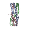
| 1 | X-RAY DIFFRACTION | 1 | (CHAIN 'A' AND RESID 4 THROUGH 92)| 2 | X-RAY DIFFRACTION | 2 | (CHAIN 'B' AND RESID 4 THROUGH 92)| 3 | X-RAY DIFFRACTION | 3 | (CHAIN 'C' AND RESID 4 THROUGH 92) | | |
|
|---|
 ムービー
ムービー コントローラー


 データを開く
 基本情報
基本情報 要素
要素 キーワード
キーワード 機能・相同性情報
 データ登録者
 米国, 2件
  引用
引用 構造の表示
構造の表示 ダウンロードとリンク
 その他のダウンロード
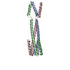


 PDBj
PDBj


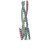
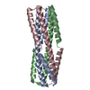
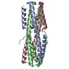
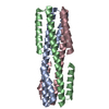
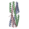
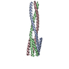
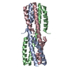
 集合体
集合体


 分子量: 79.904 Da / 分子数: 2 / 由来タイプ: 合成 / 式: Br
 分子量: 18.015 Da / 分子数: 11 / 由来タイプ: 天然 / 式: H2O
 試料調製
試料調製 解析
解析