 Movie
Movie Controller


+ Open data

- Basic information
| Entry | Database: EMDB / ID: EMD-30622 | |||||||||
|---|---|---|---|---|---|---|---|---|---|---|
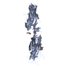
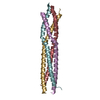
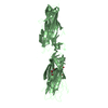
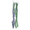
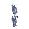
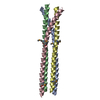
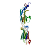
| Title | Cryo-EM structure of amyloid fibril formed by human RIPK3 | |||||||||
 Map data Map data | ||||||||||
 Sample Sample |
| |||||||||
 Keywords Keywords | Amyloid fibril / PROTEIN FIBRIL | |||||||||
| Function / homology |  Function and homology information Function and homology informationregulation of activation-induced cell death of T cells / regulation of CD8-positive, alpha-beta cytotoxic T cell extravasation / execution phase of necroptosis / regulation of T cell mediated cytotoxicity / ripoptosome assembly involved in necroptotic process / regulation of adaptive immune response / regulation of activated T cell proliferation / activation of protein kinase activity / Microbial modulation of RIPK1-mediated regulated necrosis / ripoptosome ...regulation of activation-induced cell death of T cells / regulation of CD8-positive, alpha-beta cytotoxic T cell extravasation / execution phase of necroptosis / regulation of T cell mediated cytotoxicity / ripoptosome assembly involved in necroptotic process / regulation of adaptive immune response / regulation of activated T cell proliferation / activation of protein kinase activity / Microbial modulation of RIPK1-mediated regulated necrosis / ripoptosome / TRIF-mediated programmed cell death / regulation of type II interferon production / TLR3-mediated TICAM1-dependent programmed cell death / programmed necrotic cell death / SARS-CoV-1-mediated effects on programmed cell death / necroptotic signaling pathway / RIP-mediated NFkB activation via ZBP1 / positive regulation of necroptotic process / RIPK1-mediated regulated necrosis / non-canonical NF-kappaB signal transduction / TRP channels / necroptotic process / T cell homeostasis / lymph node development / spleen development / positive regulation of intrinsic apoptotic signaling pathway / reactive oxygen species metabolic process / thymus development / : / protein modification process / TICAM1, RIP1-mediated IKK complex recruitment / protein serine/threonine kinase binding / IKK complex recruitment mediated by RIP1 / apoptotic signaling pathway / Regulation of necroptotic cell death / positive regulation of reactive oxygen species metabolic process / cellular response to hydrogen peroxide / SARS-CoV-1 activates/modulates innate immune responses / T cell differentiation in thymus / regulation of apoptotic process / defense response to virus / amyloid fibril formation / protein kinase activity / transcription coactivator activity / non-specific serine/threonine protein kinase / protein serine kinase activity / protein serine/threonine kinase activity / protein-containing complex binding / signal transduction / protein-containing complex / ATP binding / identical protein binding / nucleus / cytoplasm / cytosol Similarity search - Function | |||||||||
| Biological species |  Homo sapiens (human) Homo sapiens (human) | |||||||||
| Method | helical reconstruction / cryo EM / Resolution: 4.24 Å | |||||||||
 Authors Authors | Zhao K / Ma YY | |||||||||
 Citation Citation | Journal: Proc Natl Acad Sci U S A / Year: 2021 Title: The structure of a minimum amyloid fibril core formed by necroptosis-mediating RHIM of human RIPK3. Authors: Xialian Wu / Yeyang Ma / Kun Zhao / Jing Zhang / Yunpeng Sun / Yichen Li / Xingqi Dong / Hong Hu / Jing Liu / Jian Wang / Xia Zhang / Bing Li / Huayi Wang / Dan Li / Bo Sun / Junxia Lu / Cong Liu /  Abstract: Receptor-interacting protein kinases 3 (RIPK3), a central node in necroptosis, polymerizes in response to the upstream signals and then activates its downstream mediator to induce cell death. The ...Receptor-interacting protein kinases 3 (RIPK3), a central node in necroptosis, polymerizes in response to the upstream signals and then activates its downstream mediator to induce cell death. The active polymeric form of RIPK3 has been indicated as the form of amyloid fibrils assembled via its RIP homotypic interaction motif (RHIM). In this study, we combine cryogenic electron microscopy and solid-state NMR to determine the amyloid fibril structure of RIPK3 RHIM-containing C-terminal domain (CTD). The structure reveals a single protofilament composed of the RHIM domain. RHIM forms three β-strands (referred to as strands 1 through 3) folding into an S shape, a distinct fold from that in complex with RIPK1. The consensus tetrapeptide VQVG of RHIM forms strand 2, which zips up strands 1 and 3 via heterozipper-like interfaces. Notably, the RIPK3-CTD fibril, as a physiological fibril, exhibits distinctive assembly compared with pathological fibrils. It has an exceptionally small fibril core and twists in both handedness with the smallest pitch known so far. These traits may contribute to a favorable spatial arrangement of RIPK3 kinase domain for efficient phosphorylation. | |||||||||
| History |
|
- Structure visualization
| Movie |
Movie viewer |
|---|---|
| Structure viewer | EM map: SurfViewMolmilJmol/JSmol |
| Supplemental images |

- Downloads & links
-EMDB archive
| Map data | emd_30622.map.gz | 5.1 MB | EMDB map data format | |
|---|---|---|---|---|
| Header (meta data) | emd-30622-v30.xmlemd-30622.xml | 9.4 KB 9.4 KB | Display Display | EMDB header |
| FSC (resolution estimation) | emd_30622_fsc.xml | 10.3 KB | Display | FSC data file |
| Images |  emd_30622.png emd_30622.png | 32.6 KB | ||
| Filedesc metadata | emd-30622.cif.gz | 4.8 KB | ||
| Archive directory |  http://ftp.pdbj.org/pub/emdb/structures/EMD-30622ftp://ftp.pdbj.org/pub/emdb/structures/EMD-30622 http://ftp.pdbj.org/pub/emdb/structures/EMD-30622ftp://ftp.pdbj.org/pub/emdb/structures/EMD-30622 | HTTPS FTP |
-Related structure data
| Related structure data | 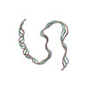 7da4MC 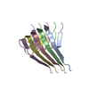 7dacC M: atomic model generated by this map C: citing same article ( |
|---|---|
| Similar structure data |


-Links
| EMDB pages | EMDB (EBI/PDBe) / EMDataResource |
|---|---|
| Related items in Molecule of the Month |


-Map
| File | Download / File: emd_30622.map.gz / Format: CCP4 / Size: 91.1 MB / Type: IMAGE STORED AS FLOATING POINT NUMBER (4 BYTES) | ||||||||||||||||||||||||||||||||||||||||||||||||||||||||||||||||||||
|---|---|---|---|---|---|---|---|---|---|---|---|---|---|---|---|---|---|---|---|---|---|---|---|---|---|---|---|---|---|---|---|---|---|---|---|---|---|---|---|---|---|---|---|---|---|---|---|---|---|---|---|---|---|---|---|---|---|---|---|---|---|---|---|---|---|---|---|---|---|
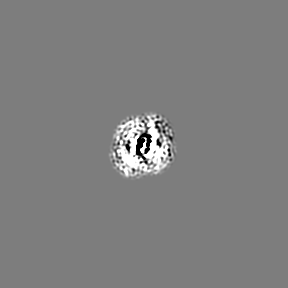
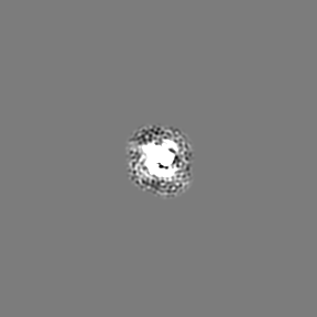
| Projections & slices | Image control
Images are generated by Spider. | ||||||||||||||||||||||||||||||||||||||||||||||||||||||||||||||||||||
| Voxel size | X=Y=Z: 1.06 Å | ||||||||||||||||||||||||||||||||||||||||||||||||||||||||||||||||||||
| Density |
| ||||||||||||||||||||||||||||||||||||||||||||||||||||||||||||||||||||
| Symmetry | Space group: 1 | ||||||||||||||||||||||||||||||||||||||||||||||||||||||||||||||||||||
| Details | EMDB XML:
CCP4 map header:
| ||||||||||||||||||||||||||||||||||||||||||||||||||||||||||||||||||||
 Z (Sec.)
 Y (Row.)
 X (Col.)


-Supplemental data
- Sample components
-Entire : human RIPK3-CTD amyloid fibril
| Entire | Name: human RIPK3-CTD amyloid fibril |
|---|---|
| Components |
|
-Supramolecule #1: human RIPK3-CTD amyloid fibril
| Supramolecule | Name: human RIPK3-CTD amyloid fibril / type: organelle_or_cellular_component / ID: 1 / Parent: 0 / Macromolecule list: all |
|---|---|
| Source (natural) | Organism: Homo sapiens (human) |
-Macromolecule #1: Receptor-interacting serine/threonine-protein kinase 3
| Macromolecule | Name: Receptor-interacting serine/threonine-protein kinase 3 type: protein_or_peptide / ID: 1 / Number of copies: 3 / Enantiomer: LEVO / EC number: non-specific serine/threonine protein kinase |
|---|---|
| Source (natural) | Organism: Homo sapiens (human) |
| Molecular weight | Theoretical: 15.311755 KDa |
| Recombinant expression | Organism:  |
| Sequence | String: MGSSDSMAQP PQTPETSTFR NQMPSPTSTG TPSPGPRGNQ GAERQGMNWS CRTPEPNPVT GRPLVNIYNC SGVQVGDNNY LTMQQTTAL PTWGLAPSGK GRGLQHPPPV GSQEGPKDPE AWSRPQGWYN HSGKLEHHHH HH UniProtKB: Receptor-interacting serine/threonine-protein kinase 3 |
-Experimental details
-Structure determination
| Method | cryo EM |
|---|---|
 Processing Processing | helical reconstruction |
| Aggregation state | helical array |
-Sample preparation
| Buffer | pH: 5 |
|---|---|
| Vitrification | Cryogen name: ETHANE |
- Electron microscopy
| Microscope | FEI TITAN KRIOS |
|---|---|
| Image recording | Film or detector model: GATAN K3 BIOQUANTUM (6k x 4k) / Average electron dose: 55.0 e/Å2 |
| Electron beam | Acceleration voltage: 300 kV / Electron source:  FIELD EMISSION GUN FIELD EMISSION GUN |
| Electron optics | Illumination mode: FLOOD BEAM / Imaging mode: BRIGHT FIELD |
| Experimental equipment |  Model: Titan Krios / Image courtesy: FEI Company |
-Image processing
| Final reconstruction | Applied symmetry - Helical parameters - Δz: 4.8 Å Applied symmetry - Helical parameters - Δ&Phi: -7.53 ° Applied symmetry - Helical parameters - Axial symmetry: C1 (asymmetric) Resolution.type: BY AUTHOR / Resolution: 4.24 Å / Resolution method: FSC 0.143 CUT-OFF / Number images used: 45526 |
|---|---|
| Startup model | Type of model: NONE |
| Final angle assignment | Type: NOT APPLICABLE |
| FSC plot (resolution estimation) |  |
