 Movie
Movie Controller


+ Open data

- Basic information
| Entry | Database: PDB / ID: 6nxm | |||||||||
|---|---|---|---|---|---|---|---|---|---|---|
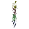
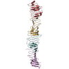
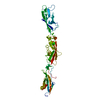
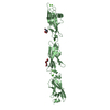
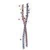
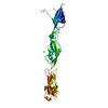
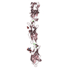
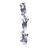
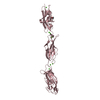
| Title | Crystal structure of computationally designed protein XAA_GVDQ | |||||||||
 Components Components | Design construct XAA_GVDQ | |||||||||
 Keywords Keywords | DE NOVO PROTEIN / homotrimer / helix | |||||||||
| Biological species | synthetic construct (others) | |||||||||
| Method |  X-RAY DIFFRACTION / SYNCHROTRON / MOLECULAR REPLACEMENT / Resolution: 2.2 Å X-RAY DIFFRACTION / SYNCHROTRON / MOLECULAR REPLACEMENT / Resolution: 2.2 Å | |||||||||
 Authors Authors | Wei, K.Y. / Bick, M.J. | |||||||||
| Funding support |  United States, 2items United States, 2items
| |||||||||
 Citation Citation | Journal: Proc.Natl.Acad.Sci.USA / Year: 2020 Title: Computational design of closely related proteins that adopt two well-defined but structurally divergent folds. Authors: Wei, K.Y. / Moschidi, D. / Bick, M.J. / Nerli, S. / McShan, A.C. / Carter, L.P. / Huang, P.S. / Fletcher, D.A. / Sgourakis, N.G. / Boyken, S.E. / Baker, D. | |||||||||
| History |
|
- Structure visualization
| Structure viewer | Molecule:  MolmilJmol/JSmol MolmilJmol/JSmol |
|---|
- Downloads & links
-Download
| PDBx/mmCIF format | 6nxm.cif.gz | 96.1 KB | Display | PDBx/mmCIF format |
|---|---|---|---|---|
| PDB format | pdb6nxm.ent.gz | 73.9 KB | Display | PDB format |
| PDBx/mmJSON format | 6nxm.json.gz | Tree view | PDBx/mmJSON format | |
| Others |  Other downloads Other downloads |
-Validation report
| Arichive directory | https://data.pdbj.org/pub/pdb/validation_reports/nx/6nxmftp://data.pdbj.org/pub/pdb/validation_reports/nx/6nxm | HTTPS FTP |
|---|
-Related structure data
| Related structure data | 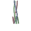 6nx2C 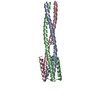 6ny8C 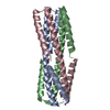 6nyeC 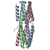 6nyiC 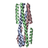 6nykC 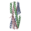 6nz1C 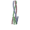 6nz3C 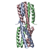 6o0cC 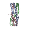 6o0iC C: citing same article ( |
|---|---|
| Similar structure data |

-Links
 PDBj
PDBj
- Assembly
| Deposited unit | 
| ||||||||
|---|---|---|---|---|---|---|---|---|---|
| 1 | 
| ||||||||
| Unit cell |
|
-Components
| #1: Protein | Mass: 10991.570 Da / Num. of mol.: 3 Source method: isolated from a genetically manipulated source Source: (gene. exp.) synthetic construct (others) / Plasmid: pET28b / Production host:  #2: Water | ChemComp-HOH / |  Mass: 18.015 Da / Num. of mol.: 6 / Source method: isolated from a natural source / Formula: H2O Mass: 18.015 Da / Num. of mol.: 6 / Source method: isolated from a natural source / Formula: H2O |
|---|
-Experimental details
-Experiment
| Experiment | Method: X-RAY DIFFRACTION / Number of used crystals: 1 |
|---|
- Sample preparation
| Crystal | Density Matthews: 2.15 Å3/Da / Density % sol: 42.76 % |
|---|---|
| Crystal grow | Temperature: 291 K / Method: vapor diffusion, sitting drop / pH: 4.6 / Details: 2.0 M sodium formate, 0.1 M sodium acetate pH 4.6 |
-Data collection
| Diffraction | Mean temperature: 100 K / Serial crystal experiment: N |
|---|---|
| Diffraction source | Source: SYNCHROTRON / Site: ALS / Beamline: 8.3.1 / Wavelength: 1.1111 Å |
| Detector | Type: DECTRIS PILATUS3 S 6M / Detector: PIXEL / Date: Oct 19, 2017 |
| Radiation | Protocol: SINGLE WAVELENGTH / Monochromatic (M) / Laue (L): M / Scattering type: x-ray |
| Radiation wavelength | Wavelength: 1.1111 Å / Relative weight: 1 |
| Reflection | Resolution: 1.82→48.133 Å / Num. obs: 23257 / % possible obs: 92.44 % / Redundancy: 6.3 % / Rmerge(I) obs: 0.041 / Rpim(I) all: 0.018 / Rrim(I) all: 0.045 / Net I/σ(I): 14.1 |
| Reflection shell | Resolution: 1.82→1.89 Å / Num. unique obs: 1365 / CC1/2: 0.011 / % possible all: 54.62 |
- Processing
| Software |
| ||||||||||||||||||||||||||||||||||||||||||||||||||||||||||||||||||||||
|---|---|---|---|---|---|---|---|---|---|---|---|---|---|---|---|---|---|---|---|---|---|---|---|---|---|---|---|---|---|---|---|---|---|---|---|---|---|---|---|---|---|---|---|---|---|---|---|---|---|---|---|---|---|---|---|---|---|---|---|---|---|---|---|---|---|---|---|---|---|---|---|
| Refinement | Method to determine structure: MOLECULAR REPLACEMENT / Resolution: 2.2→48.133 Å / SU ML: 0.39 / Cross valid method: THROUGHOUT / σ(F): 0 / Phase error: 36.84
| ||||||||||||||||||||||||||||||||||||||||||||||||||||||||||||||||||||||
| Solvent computation | Shrinkage radii: 0.9 Å / VDW probe radii: 1.11 Å | ||||||||||||||||||||||||||||||||||||||||||||||||||||||||||||||||||||||
| Displacement parameters | Biso max: 166.42 Å2 / Biso mean: 78.4719 Å2 / Biso min: 39.22 Å2 | ||||||||||||||||||||||||||||||||||||||||||||||||||||||||||||||||||||||
| Refinement step | Cycle: final / Resolution: 2.2→48.133 Å
| ||||||||||||||||||||||||||||||||||||||||||||||||||||||||||||||||||||||
| LS refinement shell | Refine-ID: X-RAY DIFFRACTION / Rfactor Rfree error: 0 / Total num. of bins used: 9
|
