+ Open data
- Basic information
| Entry | Database: PDB / ID: 6nuu | |||||||||
|---|---|---|---|---|---|---|---|---|---|---|
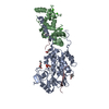
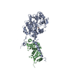
| Title | Structure of Calcineurin mutant in complex with NHE1 peptide | |||||||||
 Components Components |
| |||||||||
 Keywords Keywords | HYDROLASE/Calcium Binding Protein / Ser/thr phosphatase / complex / HYDROLASE / HYDROLASE-Calcium Binding Protein complex | |||||||||
| Function / homology |  Function and homology information Function and homology informationcation-transporting ATPase complex / Sodium/Proton exchangers / negative regulation of angiotensin-activated signaling pathway / calcium-dependent protein serine/threonine phosphatase regulator activity / regulation of cell proliferation involved in kidney morphogenesis / positive regulation of glomerulus development / negative regulation of calcium ion import across plasma membrane / regulation of the force of heart contraction by cardiac conduction / negative regulation of signaling / calcium-dependent protein serine/threonine phosphatase activity ...cation-transporting ATPase complex / Sodium/Proton exchangers / negative regulation of angiotensin-activated signaling pathway / calcium-dependent protein serine/threonine phosphatase regulator activity / regulation of cell proliferation involved in kidney morphogenesis / positive regulation of glomerulus development / negative regulation of calcium ion import across plasma membrane / regulation of the force of heart contraction by cardiac conduction / negative regulation of signaling / calcium-dependent protein serine/threonine phosphatase activity / Hyaluronan degradation / positive regulation of saliva secretion / peptidyl-serine dephosphorylation / regulation of cardiac muscle cell membrane potential / calmodulin-dependent protein phosphatase activity / calcineurin complex / positive regulation of connective tissue replacement / positive regulation of calcium ion import across plasma membrane / positive regulation of calcium ion-dependent exocytosis of neurotransmitter / positive regulation of cardiac muscle hypertrophy in response to stress / cellular response to electrical stimulus / negative regulation of dendrite morphogenesis / protein serine/threonine phosphatase complex / potassium:proton antiporter activity / renal filtration / positive regulation of action potential / lung epithelial cell differentiation / sodium:proton antiporter activity / calcineurin-NFAT signaling cascade / maintenance of cell polarity / regulation of pH / sodium ion export across plasma membrane / positive regulation of calcineurin-NFAT signaling cascade / cardiac muscle cell differentiation / skeletal muscle tissue regeneration / myelination in peripheral nervous system / response to acidic pH / protein phosphatase 2B binding / transition between fast and slow fiber / intracellular sodium ion homeostasis / regulation of stress fiber assembly / cellular response to acidic pH / positive regulation of osteoclast differentiation / sodium ion import across plasma membrane / cardiac muscle hypertrophy in response to stress / cardiac muscle cell contraction / regulation of synaptic vesicle cycle / positive regulation of mitochondrial membrane permeability / dephosphorylation / regulation of cardiac muscle contraction by calcium ion signaling / regulation of focal adhesion assembly / cellular response to antibiotic / extrinsic component of plasma membrane / CLEC7A (Dectin-1) induces NFAT activation / branching involved in blood vessel morphogenesis / dendrite morphogenesis / positive regulation of cardiac muscle hypertrophy / cellular response to cold / protein-serine/threonine phosphatase / regulation of postsynaptic neurotransmitter receptor internalization / parallel fiber to Purkinje cell synapse / positive regulation of the force of heart contraction / calcineurin-mediated signaling / protein serine/threonine phosphatase activity / positive regulation of activated T cell proliferation / protein complex oligomerization / positive regulation of endocytosis / Calcineurin activates NFAT / intercalated disc / DARPP-32 events / epithelial to mesenchymal transition / epidermis development / Activation of BAD and translocation to mitochondria / positive regulation of osteoblast differentiation / multicellular organismal response to stress / phosphatase binding / postsynaptic modulation of chemical synaptic transmission / protein dephosphorylation / skeletal muscle fiber development / keratinocyte differentiation / monoatomic ion transport / response to muscle stretch / phosphatidylinositol-4,5-bisphosphate binding / cellular response to epinephrine stimulus / potassium ion transmembrane transport / T-tubule / FCERI mediated Ca+2 mobilization / proton transmembrane transport / positive regulation of cell adhesion / T cell activation / hippocampal mossy fiber to CA3 synapse / excitatory postsynaptic potential / regulation of intracellular pH / stem cell differentiation / wound healing / cellular response to mechanical stimulus / G1/S transition of mitotic cell cycle / response to calcium ion / sarcolemma / modulation of chemical synaptic transmission Similarity search - Function | |||||||||
| Biological species |  Homo sapiens (human) Homo sapiens (human) | |||||||||
| Method |  X-RAY DIFFRACTION / X-RAY DIFFRACTION /  MOLECULAR REPLACEMENT / Resolution: 2.3 Å MOLECULAR REPLACEMENT / Resolution: 2.3 Å | |||||||||
 Authors Authors | Wang, X. / Page, R. / Peti, W. | |||||||||
| Funding support |  United States, 2items United States, 2items
| |||||||||
 Citation Citation |  Journal: Nat Commun / Year: 2019 Journal: Nat Commun / Year: 2019Title: Molecular basis for the binding and selective dephosphorylation of Na+/H+exchanger 1 by calcineurin. Authors: Hendus-Altenburger, R. / Wang, X. / Sjogaard-Frich, L.M. / Pedraz-Cuesta, E. / Sheftic, S.R. / Bendsoe, A.H. / Page, R. / Kragelund, B.B. / Pedersen, S.F. / Peti, W. | |||||||||
| History |
|
- Structure visualization
| Structure viewer | Molecule:  Molmil Molmil Jmol/JSmol Jmol/JSmol |
|---|
- Downloads & links
- Download
| PDBx/mmCIF format |  6nuu.cif.gz 6nuu.cif.gz | 220.8 KB | Display |  PDBx/mmCIF format PDBx/mmCIF format |
|---|---|---|---|---|
| PDB format |  pdb6nuu.ent.gz pdb6nuu.ent.gz | 176 KB | Display |  PDB format PDB format |
| PDBx/mmJSON format |  6nuu.json.gz 6nuu.json.gz | Tree view |  PDBx/mmJSON format PDBx/mmJSON format | |
| Others |  Other downloads Other downloads |
-Validation report
| Summary document |  6nuu_validation.pdf.gz 6nuu_validation.pdf.gz | 462.9 KB | Display |  wwPDB validaton report wwPDB validaton report |
|---|---|---|---|---|
| Full document |  6nuu_full_validation.pdf.gz 6nuu_full_validation.pdf.gz | 465.3 KB | Display | |
| Data in XML |  6nuu_validation.xml.gz 6nuu_validation.xml.gz | 22.9 KB | Display | |
| Data in CIF |  6nuu_validation.cif.gz 6nuu_validation.cif.gz | 32.8 KB | Display | |
| Arichive directory |  https://data.pdbj.org/pub/pdb/validation_reports/nu/6nuu https://data.pdbj.org/pub/pdb/validation_reports/nu/6nuu ftp://data.pdbj.org/pub/pdb/validation_reports/nu/6nuu ftp://data.pdbj.org/pub/pdb/validation_reports/nu/6nuu | HTTPS FTP |
-Related structure data
| Related structure data | 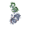 6nucC  6nufC  5sveS S: Starting model for refinement C: citing same article ( |
|---|---|
| Similar structure data |
- Links
Links
- Assembly
| Deposited unit | 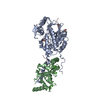
| |||||||||||||||
|---|---|---|---|---|---|---|---|---|---|---|---|---|---|---|---|---|
| 1 |
| |||||||||||||||
| Unit cell |
| |||||||||||||||
| Components on special symmetry positions |
|
- Components
-Protein , 2 types, 2 molecules AB
| #1: Protein | Mass: 42695.883 Da / Num. of mol.: 1 / Mutation: H155A, Y159A Source method: isolated from a genetically manipulated source Source: (gene. exp.)  Homo sapiens (human) / Gene: PPP3CA, CALNA, CNA / Production host: Homo sapiens (human) / Gene: PPP3CA, CALNA, CNA / Production host:  References: UniProt: Q08209, protein-serine/threonine phosphatase |
|---|---|
| #2: Protein | Mass: 17755.174 Da / Num. of mol.: 1 Source method: isolated from a genetically manipulated source Source: (gene. exp.)  Homo sapiens (human) / Gene: PPP3R1, CNA2, CNB / Production host: Homo sapiens (human) / Gene: PPP3R1, CNA2, CNB / Production host:  |
-Protein/peptide , 1 types, 1 molecules C
| #3: Protein/peptide | Mass: 5268.008 Da / Num. of mol.: 1 Source method: isolated from a genetically manipulated source Source: (gene. exp.)  Homo sapiens (human) / Gene: SLC9A1, APNH1, NHE1 / Production host: Homo sapiens (human) / Gene: SLC9A1, APNH1, NHE1 / Production host:  |
|---|
-Non-polymers , 6 types, 264 molecules 




| #4: Chemical | ChemComp-FE / | ||
|---|---|---|---|
| #5: Chemical | ChemComp-ZN / | ||
| #6: Chemical | ChemComp-PO4 / | ||
| #7: Chemical | ChemComp-PEG / | ||
| #8: Chemical | ChemComp-CA / #9: Water | ChemComp-HOH / | |
-Experimental details
-Experiment
| Experiment | Method:  X-RAY DIFFRACTION / Number of used crystals: 1 X-RAY DIFFRACTION / Number of used crystals: 1 |
|---|
- Sample preparation
| Crystal | Density Matthews: 2.49 Å3/Da / Density % sol: 50.6 % |
|---|---|
| Crystal grow | Temperature: 298 K / Method: vapor diffusion, sitting drop / pH: 9.5 Details: 40% (v/v) PEG 600, 100 mM CHES/ Sodium hydroxide pH 9.5, 0.2 M MgCl2 |
-Data collection
| Diffraction | Mean temperature: 100 K / Serial crystal experiment: N |
|---|---|
| Diffraction source | Source: LIQUID ANODE / Type: BRUKER METALJET / Wavelength: 1.34 Å |
| Detector | Type: Bruker PHOTON II / Detector: PIXEL / Date: Nov 18, 2018 |
| Radiation | Protocol: SINGLE WAVELENGTH / Monochromatic (M) / Laue (L): M / Scattering type: x-ray |
| Radiation wavelength | Wavelength: 1.34 Å / Relative weight: 1 |
| Reflection | Resolution: 2.3→20.8 Å / Num. obs: 28153 / % possible obs: 99.8 % / Redundancy: 5.3 % / CC1/2: 0.984 / Net I/σ(I): 5.9 |
| Reflection shell | Resolution: 2.3→2.38 Å / Num. unique obs: 14368 / CC1/2: 0.714 |
- Processing
| Software |
| |||||||||||||||||||||||||||||||||||||||||||||||||||||||||||||||||||||||||||||||||||||||||||||||||||||||||
|---|---|---|---|---|---|---|---|---|---|---|---|---|---|---|---|---|---|---|---|---|---|---|---|---|---|---|---|---|---|---|---|---|---|---|---|---|---|---|---|---|---|---|---|---|---|---|---|---|---|---|---|---|---|---|---|---|---|---|---|---|---|---|---|---|---|---|---|---|---|---|---|---|---|---|---|---|---|---|---|---|---|---|---|---|---|---|---|---|---|---|---|---|---|---|---|---|---|---|---|---|---|---|---|---|---|---|
| Refinement | Method to determine structure:  MOLECULAR REPLACEMENT MOLECULAR REPLACEMENTStarting model: 5SVE Resolution: 2.3→20.8 Å / SU ML: 0.25 / Cross valid method: FREE R-VALUE / σ(F): 0 / Phase error: 24.69
| |||||||||||||||||||||||||||||||||||||||||||||||||||||||||||||||||||||||||||||||||||||||||||||||||||||||||
| Solvent computation | Shrinkage radii: 0.9 Å / VDW probe radii: 1.11 Å | |||||||||||||||||||||||||||||||||||||||||||||||||||||||||||||||||||||||||||||||||||||||||||||||||||||||||
| Refinement step | Cycle: LAST / Resolution: 2.3→20.8 Å
| |||||||||||||||||||||||||||||||||||||||||||||||||||||||||||||||||||||||||||||||||||||||||||||||||||||||||
| Refine LS restraints |
| |||||||||||||||||||||||||||||||||||||||||||||||||||||||||||||||||||||||||||||||||||||||||||||||||||||||||
| LS refinement shell |
|
 Movie
Movie Controller



 PDBj
PDBj


