 Movie
Movie Controller

[English] 日本語
 Yorodumi
- PDB-6nt8: Cryo-EM structure of full-length chicken STING in the cGAMP-bound... -
+ Open data

- Basic information
| Entry | Database: PDB / ID: 6nt8 | |||||||||||||||||||||
|---|---|---|---|---|---|---|---|---|---|---|---|---|---|---|---|---|---|---|---|---|---|---|
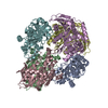
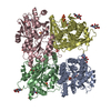
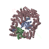
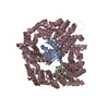
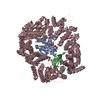
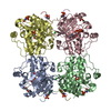
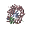
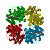
| Title | Cryo-EM structure of full-length chicken STING in the cGAMP-bound tetrameric state | |||||||||||||||||||||
 Components Components | Stimulator of interferon genes protein | |||||||||||||||||||||
 Keywords Keywords | IMMUNE SYSTEM / ER / membrane / adaptor | |||||||||||||||||||||
| Function / homology |  Function and homology information Function and homology informationSTING mediated induction of host immune responses / 2',3'-cyclic GMP-AMP binding / cyclic-di-GMP binding / proton channel activity / reticulophagy / cGAS/STING signaling pathway / protein complex oligomerization / autophagosome membrane / positive regulation of macroautophagy / autophagosome assembly ...STING mediated induction of host immune responses / 2',3'-cyclic GMP-AMP binding / cyclic-di-GMP binding / proton channel activity / reticulophagy / cGAS/STING signaling pathway / protein complex oligomerization / autophagosome membrane / positive regulation of macroautophagy / autophagosome assembly / positive regulation of type I interferon production / endoplasmic reticulum-Golgi intermediate compartment membrane / activation of innate immune response / positive regulation of interferon-beta production / autophagosome / Neutrophil degranulation / cytoplasmic vesicle / defense response to virus / Golgi membrane / innate immune response / endoplasmic reticulum membrane / perinuclear region of cytoplasm / protein homodimerization activity / cytoplasm Similarity search - Function | |||||||||||||||||||||
| Biological species |  | |||||||||||||||||||||
| Method | ELECTRON MICROSCOPY / single particle reconstruction / cryo EM / Resolution: 6.5 Å | |||||||||||||||||||||
 Authors Authors | Shang, G. / Zhang, C. / Chen, Z.J. / Bai, X. / Zhang, X. | |||||||||||||||||||||
| Funding support |  United States, 6items United States, 6items
| |||||||||||||||||||||
 Citation Citation | Journal: Nature / Year: 2019 Title: Cryo-EM structures of STING reveal its mechanism of activation by cyclic GMP-AMP. Authors: Guijun Shang / Conggang Zhang / Zhijian J Chen / Xiao-Chen Bai / Xuewu Zhang / Abstract: Infections by pathogens that contain DNA trigger the production of type-I interferons and inflammatory cytokines through cyclic GMP-AMP synthase, which produces 2'3'-cyclic GMP-AMP (cGAMP) that binds ...Infections by pathogens that contain DNA trigger the production of type-I interferons and inflammatory cytokines through cyclic GMP-AMP synthase, which produces 2'3'-cyclic GMP-AMP (cGAMP) that binds to and activates stimulator of interferon genes (STING; also known as TMEM173, MITA, ERIS and MPYS). STING is an endoplasmic-reticulum membrane protein that contains four transmembrane helices followed by a cytoplasmic ligand-binding and signalling domain. The cytoplasmic domain of STING forms a dimer, which undergoes a conformational change upon binding to cGAMP. However, it remains unclear how this conformational change leads to STING activation. Here we present cryo-electron microscopy structures of full-length STING from human and chicken in the inactive dimeric state (about 80 kDa in size), as well as cGAMP-bound chicken STING in both the dimeric and tetrameric states. The structures show that the transmembrane and cytoplasmic regions interact to form an integrated, domain-swapped dimeric assembly. Closure of the ligand-binding domain, induced by cGAMP, leads to a 180° rotation of the ligand-binding domain relative to the transmembrane domain. This rotation is coupled to a conformational change in a loop on the side of the ligand-binding-domain dimer, which leads to the formation of the STING tetramer and higher-order oligomers through side-by-side packing. This model of STING oligomerization and activation is supported by our structure-based mutational analyses. | |||||||||||||||||||||
| History |
|
- Structure visualization
| Movie |
Movie viewer |
|---|---|
| Structure viewer | Molecule: MolmilJmol/JSmol |
- Downloads & links
-Download
| PDBx/mmCIF format | 6nt8.cif.gz | 222.1 KB | Display | PDBx/mmCIF format |
|---|---|---|---|---|
| PDB format | pdb6nt8.ent.gz | 175.5 KB | Display | PDB format |
| PDBx/mmJSON format | 6nt8.json.gz | Tree view | PDBx/mmJSON format | |
| Others |  Other downloads Other downloads |
-Validation report
| Arichive directory | https://data.pdbj.org/pub/pdb/validation_reports/nt/6nt8ftp://data.pdbj.org/pub/pdb/validation_reports/nt/6nt8 | HTTPS FTP |
|---|
-Related structure data
| Related structure data | 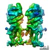 0505MC 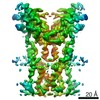 0502C 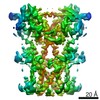 0503C 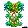 0504C 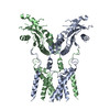 6nt5C 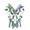 6nt6C 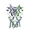 6nt7C M: map data used to model this data C: citing same article ( |
|---|---|
| Similar structure data |
-Links
 PDBj
PDBj- Assembly
| Deposited unit | 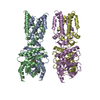
|
|---|---|
| 1 |
|
-Components
| #1: Protein | Mass: 44207.070 Da / Num. of mol.: 4 Source method: isolated from a genetically manipulated source Source: (gene. exp.)  Homo sapiens (human) / References: UniProt: A0A1D5P7Q9, UniProt: E1C7U0*PLUS Homo sapiens (human) / References: UniProt: A0A1D5P7Q9, UniProt: E1C7U0*PLUS#2: Chemical |   Mass: 674.411 Da / Num. of mol.: 2 / Source method: obtained synthetically / Formula: C20H24N10O13P2 / Feature type: SUBJECT OF INVESTIGATION Mass: 674.411 Da / Num. of mol.: 2 / Source method: obtained synthetically / Formula: C20H24N10O13P2 / Feature type: SUBJECT OF INVESTIGATION |
|---|
-Experimental details
-Experiment
| Experiment | Method: ELECTRON MICROSCOPY |
|---|---|
| EM experiment | Aggregation state: PARTICLE / 3D reconstruction method: single particle reconstruction |
- Sample preparation
| Component | Name: full-length chicken STING / Type: COMPLEX / Entity ID: #1 / Source: RECOMBINANT |
|---|---|
| Source (natural) | Organism: |
| Source (recombinant) | Organism: Homo sapiens (human) / Cell: HEK293 GnTI- |
| Buffer solution | pH: 8 |
| Specimen | Conc.: 4.5 mg/ml / Embedding applied: NO / Shadowing applied: NO / Staining applied: NO / Vitrification applied: YES |
| Specimen support | Details: unspecified |
| Vitrification | Cryogen name: ETHANE |
- Electron microscopy imaging
| Experimental equipment |  Model: Titan Krios / Image courtesy: FEI Company |
|---|---|
| Microscopy | Model: FEI TITAN KRIOS |
| Electron gun | Electron source:  FIELD EMISSION GUN / Accelerating voltage: 300 kV / Illumination mode: FLOOD BEAM FIELD EMISSION GUN / Accelerating voltage: 300 kV / Illumination mode: FLOOD BEAM |
| Electron lens | Mode: BRIGHT FIELD |
| Specimen holder | Cryogen: NITROGEN / Specimen holder model: FEI TITAN KRIOS AUTOGRID HOLDER |
| Image recording | Electron dose: 40 e/Å2 / Detector mode: SUPER-RESOLUTION / Film or detector model: GATAN K2 SUMMIT (4k x 4k) |
| Image scans | Movie frames/image: 20 |
- Processing
| EM software |
| |||||||||||||||||||||||||||
|---|---|---|---|---|---|---|---|---|---|---|---|---|---|---|---|---|---|---|---|---|---|---|---|---|---|---|---|---|
| CTF correction | Type: PHASE FLIPPING AND AMPLITUDE CORRECTION | |||||||||||||||||||||||||||
| Symmetry | Point symmetry: C1 (asymmetric) | |||||||||||||||||||||||||||
| 3D reconstruction | Resolution: 6.5 Å / Resolution method: FSC 0.143 CUT-OFF / Num. of particles: 41033 / Symmetry type: POINT | |||||||||||||||||||||||||||
| Atomic model building | Protocol: RIGID BODY FIT / Space: REAL |
