 Movie
Movie Controller

[English] 日本語
 Yorodumi
- PDB-6npf: Structure of E.coli enolase in complex with an analog of the natu... -
+ Open data

- Basic information
| Entry | Database: PDB / ID: 6npf | ||||||
|---|---|---|---|---|---|---|---|
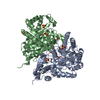
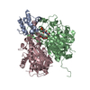
| Title | Structure of E.coli enolase in complex with an analog of the natural product SF-2312 metabolite. | ||||||
 Components Components | Enolase | ||||||
 Keywords Keywords | LYASE / Natural inhibitor / complex / enolase / SF2312 | ||||||
| Function / homology |  Function and homology information Function and homology informationphosphopyruvate hydratase / phosphopyruvate hydratase complex / phosphopyruvate hydratase activity / RNA catabolic process / RNA processing / glycolytic process / cytoskeleton / magnesium ion binding / cell surface / protein homodimerization activity ...phosphopyruvate hydratase / phosphopyruvate hydratase complex / phosphopyruvate hydratase activity / RNA catabolic process / RNA processing / glycolytic process / cytoskeleton / magnesium ion binding / cell surface / protein homodimerization activity / extracellular region / membrane / identical protein binding / cytosol Similarity search - Function | ||||||
| Biological species |  | ||||||
| Method |  X-RAY DIFFRACTION / SYNCHROTRON / MOLECULAR REPLACEMENT / Resolution: 2.57 Å X-RAY DIFFRACTION / SYNCHROTRON / MOLECULAR REPLACEMENT / Resolution: 2.57 Å | ||||||
 Authors Authors | Erlandsen, H. / Krucinska, J. / Lombardo, M. / Wright, D. | ||||||
| Funding support |  United States, 1items United States, 1items
| ||||||
 Citation Citation | Journal: Sci Rep / Year: 2019 Title: Functional and structural basis of E. coli enolase inhibition by SF2312: a mimic of the carbanion intermediate. Authors: Krucinska, J. / Lombardo, M.N. / Erlandsen, H. / Hazeen, A. / Duay, S.S. / Pattis, J.G. / Robinson, V.L. / May, E.R. / Wright, D.L. | ||||||
| History |
|
- Structure visualization
| Structure viewer | Molecule: MolmilJmol/JSmol |
|---|
- Downloads & links
-Download
| PDBx/mmCIF format | 6npf.cif.gz | 486.8 KB | Display | PDBx/mmCIF format |
|---|---|---|---|---|
| PDB format | pdb6npf.ent.gz | 399 KB | Display | PDB format |
| PDBx/mmJSON format | 6npf.json.gz | Tree view | PDBx/mmJSON format | |
| Others |  Other downloads Other downloads |
-Validation report
| Arichive directory | https://data.pdbj.org/pub/pdb/validation_reports/np/6npfftp://data.pdbj.org/pub/pdb/validation_reports/np/6npf | HTTPS FTP |
|---|
-Related structure data
| Related structure data | 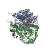 6d3qSC S: Starting model for refinement C: citing same article ( |
|---|---|
| Similar structure data |


-Links
 PDBj
PDBj

- Assembly
| Deposited unit | 
| ||||||||
|---|---|---|---|---|---|---|---|---|---|
| 1 | 
| ||||||||
| 2 | 
| ||||||||
| 3 | 
| ||||||||
| Unit cell |
|
-Components
-Protein , 1 types, 6 molecules ABCDFE
| #1: Protein | Mass: 47373.672 Da / Num. of mol.: 6 Source method: isolated from a genetically manipulated source Source: (gene. exp.) References: UniProt: B7MLA0, UniProt: P0A6P9*PLUS, phosphopyruvate hydratase |
|---|
-Non-polymers , 6 types, 276 molecules 


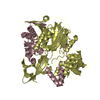
| #2: Chemical |  Mass: 150.087 Da / Num. of mol.: 3 / Source method: obtained synthetically / Formula: C4H6O6 Mass: 150.087 Da / Num. of mol.: 3 / Source method: obtained synthetically / Formula: C4H6O6#3: Chemical | ChemComp-SO4 /  Mass: 96.063 Da / Num. of mol.: 11 / Source method: obtained synthetically / Formula: SO4 Mass: 96.063 Da / Num. of mol.: 11 / Source method: obtained synthetically / Formula: SO4#4: Chemical | ChemComp-MG /  Mass: 24.305 Da / Num. of mol.: 6 / Source method: obtained synthetically / Formula: Mg Mass: 24.305 Da / Num. of mol.: 6 / Source method: obtained synthetically / Formula: Mg#5: Chemical | ChemComp-GOL /  Mass: 92.094 Da / Num. of mol.: 4 / Source method: obtained synthetically / Formula: C3H8O3 Mass: 92.094 Da / Num. of mol.: 4 / Source method: obtained synthetically / Formula: C3H8O3#6: Chemical |  Mass: 195.067 Da / Num. of mol.: 3 / Source method: obtained synthetically / Formula: C4H6NO6P / Feature type: SUBJECT OF INVESTIGATION Mass: 195.067 Da / Num. of mol.: 3 / Source method: obtained synthetically / Formula: C4H6NO6P / Feature type: SUBJECT OF INVESTIGATION#7: Water | ChemComp-HOH / | Mass: 18.015 Da / Num. of mol.: 249 / Source method: isolated from a natural source / Formula: H2O |
|---|
-Experimental details
-Experiment
| Experiment | Method: X-RAY DIFFRACTION / Number of used crystals: 1 |
|---|
- Sample preparation
| Crystal | Density Matthews: 2.26 Å3/Da / Density % sol: 45.49 % |
|---|---|
| Crystal grow | Temperature: 293 K / Method: vapor diffusion, hanging drop / pH: 6 Details: 2.1 M Ammonium Sulfate, 0.1 M MES buffer pH 6.0, 0.2 M Sodium/Potassium tartrate |
-Data collection
| Diffraction | Mean temperature: 100 K / Serial crystal experiment: N |
|---|---|
| Diffraction source | Source: SYNCHROTRON / Site: SSRL / Beamline: BL14-1 / Wavelength: 1.19499 Å |
| Detector | Type: MARMOSAIC 325 mm CCD / Detector: CCD / Date: May 10, 2018 Details: Mirror: Flat bent collimating Rh coated mirror, toroidal focussing mirror |
| Radiation | Monochromator: double crystal Si(111) / Protocol: SINGLE WAVELENGTH / Monochromatic (M) / Laue (L): M / Scattering type: x-ray |
| Radiation wavelength | Wavelength: 1.19499 Å / Relative weight: 1 |
| Reflection | Resolution: 2.57→93.03 Å / Num. obs: 97620 / % possible obs: 99.4 % / Redundancy: 1.9 % / Biso Wilson estimate: 35.5 Å2 / CC1/2: 0.984 / Rmerge(I) obs: 0.075 / Rpim(I) all: 0.075 / Rrim(I) all: 0.106 / Net I/σ(I): 5.9 |
| Reflection shell | Resolution: 2.57→2.62 Å / Redundancy: 2 % / Rmerge(I) obs: 0.396 / Mean I/σ(I) obs: 0.7 / Num. unique obs: 4421 / CC1/2: 0.791 / Rpim(I) all: 0.396 / Rrim(I) all: 0.56 / % possible all: 92.9 |
- Processing
| Software |
| ||||||||||||||||||||||||||||||||||||||||||||||||||||||||||||||||||||||||||||||||||||||||||||||||||||||||||||||||||||||||||||||||||||||||||||||||||||||||||||||||||||||||||||||||||||||
|---|---|---|---|---|---|---|---|---|---|---|---|---|---|---|---|---|---|---|---|---|---|---|---|---|---|---|---|---|---|---|---|---|---|---|---|---|---|---|---|---|---|---|---|---|---|---|---|---|---|---|---|---|---|---|---|---|---|---|---|---|---|---|---|---|---|---|---|---|---|---|---|---|---|---|---|---|---|---|---|---|---|---|---|---|---|---|---|---|---|---|---|---|---|---|---|---|---|---|---|---|---|---|---|---|---|---|---|---|---|---|---|---|---|---|---|---|---|---|---|---|---|---|---|---|---|---|---|---|---|---|---|---|---|---|---|---|---|---|---|---|---|---|---|---|---|---|---|---|---|---|---|---|---|---|---|---|---|---|---|---|---|---|---|---|---|---|---|---|---|---|---|---|---|---|---|---|---|---|---|---|---|---|---|
| Refinement | Method to determine structure: MOLECULAR REPLACEMENT Starting model: 6D3Q Resolution: 2.57→93.03 Å / Cor.coef. Fo:Fc: 0.949 / Cor.coef. Fo:Fc free: 0.895 / SU B: 14.999 / SU ML: 0.296 / Cross valid method: THROUGHOUT / ESU R: 0.599 / ESU R Free: 0.311 / Details: HYDROGENS HAVE BEEN ADDED IN THE RIDING POSITIONS
| ||||||||||||||||||||||||||||||||||||||||||||||||||||||||||||||||||||||||||||||||||||||||||||||||||||||||||||||||||||||||||||||||||||||||||||||||||||||||||||||||||||||||||||||||||||||
| Solvent computation | Ion probe radii: 0.8 Å / Shrinkage radii: 0.8 Å / VDW probe radii: 1.2 Å | ||||||||||||||||||||||||||||||||||||||||||||||||||||||||||||||||||||||||||||||||||||||||||||||||||||||||||||||||||||||||||||||||||||||||||||||||||||||||||||||||||||||||||||||||||||||
| Displacement parameters | Biso mean: 47.153 Å2
| ||||||||||||||||||||||||||||||||||||||||||||||||||||||||||||||||||||||||||||||||||||||||||||||||||||||||||||||||||||||||||||||||||||||||||||||||||||||||||||||||||||||||||||||||||||||
| Refinement step | Cycle: 1 / Resolution: 2.57→93.03 Å
| ||||||||||||||||||||||||||||||||||||||||||||||||||||||||||||||||||||||||||||||||||||||||||||||||||||||||||||||||||||||||||||||||||||||||||||||||||||||||||||||||||||||||||||||||||||||
| Refine LS restraints |
|