 Movie
Movie Controller

[English] 日本語
 Yorodumi
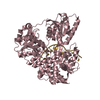
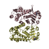
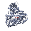
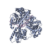
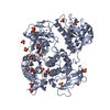
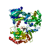
- PDB-5jaj: Structure of chicken LGP2 witha 5'p 10-mer dsRNA and ADP-AlF4-Mg. -
+ Open data

- Basic information
| Entry | Database: PDB / ID: 5jaj | ||||||
|---|---|---|---|---|---|---|---|
| Title | Structure of chicken LGP2 witha 5'p 10-mer dsRNA and ADP-AlF4-Mg. | ||||||
 Components Components |
| ||||||
 Keywords Keywords | RNA BINDING PROTEIN / Innate immune pattern recognition receptor / RIG-I like helcase / dsRNA binding / dsRNA-dependent ATPase | ||||||
| Function / homology |  Function and homology information Function and homology informationdefense response to virus / RNA helicase activity / RNA helicase / innate immune response / hydrolase activity / DNA binding / RNA binding / ATP binding / metal ion binding / cytoplasm Similarity search - Function | ||||||
| Biological species |  synthetic construct (others) | ||||||
| Method |  X-RAY DIFFRACTION / SYNCHROTRON / MOLECULAR REPLACEMENT / Resolution: 1.5 Å X-RAY DIFFRACTION / SYNCHROTRON / MOLECULAR REPLACEMENT / Resolution: 1.5 Å | ||||||
 Authors Authors | Cusack, S. / Uchikawa, E. | ||||||
| Funding support |  France, 1items France, 1items
| ||||||
 Citation Citation | Journal: Mol.Cell / Year: 2016 Title: Structural Analysis of dsRNA Binding to Anti-viral Pattern Recognition Receptors LGP2 and MDA5. Authors: Uchikawa, E. / Lethier, M. / Malet, H. / Brunel, J. / Gerlier, D. / Cusack, S. | ||||||
| History |
|
- Structure visualization
| Structure viewer | Molecule: MolmilJmol/JSmol |
|---|
- Downloads & links
-Download
| PDBx/mmCIF format | 5jaj.cif.gz | 348.6 KB | Display | PDBx/mmCIF format |
|---|---|---|---|---|
| PDB format | pdb5jaj.ent.gz | 276.2 KB | Display | PDB format |
| PDBx/mmJSON format | 5jaj.json.gz | Tree view | PDBx/mmJSON format | |
| Others |  Other downloads Other downloads |
-Validation report
| Arichive directory | https://data.pdbj.org/pub/pdb/validation_reports/ja/5jajftp://data.pdbj.org/pub/pdb/validation_reports/ja/5jaj | HTTPS FTP |
|---|
-Related structure data
| Related structure data |  5jb2C 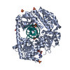 5jbgC  5jbjC 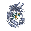 5jc3C  5jc7C  5jcfC 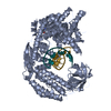 5jchC  3tmi S: Starting model for refinement C: citing same article ( |
|---|---|
| Similar structure data |


-Links
 PDBj
PDBj


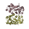
- Assembly
| Deposited unit | 
| ||||||||
|---|---|---|---|---|---|---|---|---|---|
| 1 |
| ||||||||
| Unit cell |
|
-Components
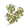
-Protein , 1 types, 1 molecules A
| #1: Protein | Mass: 77516.055 Da / Num. of mol.: 1 Mutation: GAMGGGGS at N-terminus from tag replaces methionine. Source method: isolated from a genetically manipulated source Source: (gene. exp.)  |
|---|
-RNA chain , 2 types, 2 molecules XY
| #2: RNA chain | Mass: 3482.130 Da / Num. of mol.: 1 / Source method: obtained synthetically / Source: (synth.) synthetic construct (others) |
|---|---|
| #3: RNA chain | Mass: 3176.948 Da / Num. of mol.: 1 / Source method: obtained synthetically / Source: (synth.) synthetic construct (others) |
-Non-polymers , 7 types, 571 molecules 


| #4: Chemical | ChemComp-ZN /  Mass: 65.409 Da / Num. of mol.: 1 / Source method: obtained synthetically / Formula: Zn Mass: 65.409 Da / Num. of mol.: 1 / Source method: obtained synthetically / Formula: Zn | ||||
|---|---|---|---|---|---|
| #5: Chemical | ChemComp-ADP /  Mass: 427.201 Da / Num. of mol.: 1 / Source method: obtained synthetically / Formula: C10H15N5O10P2 / Comment: ADP, energy-carrying molecule*YM Mass: 427.201 Da / Num. of mol.: 1 / Source method: obtained synthetically / Formula: C10H15N5O10P2 / Comment: ADP, energy-carrying molecule*YM | ||||
| #6: Chemical | ChemComp-ALF /  Mass: 102.975 Da / Num. of mol.: 1 / Source method: obtained synthetically / Formula: AlF4 Mass: 102.975 Da / Num. of mol.: 1 / Source method: obtained synthetically / Formula: AlF4 | ||||
| #7: Chemical | ChemComp-MG /  Mass: 24.305 Da / Num. of mol.: 1 / Source method: obtained synthetically / Formula: Mg Mass: 24.305 Da / Num. of mol.: 1 / Source method: obtained synthetically / Formula: Mg | ||||
| #8: Chemical |  Mass: 62.068 Da / Num. of mol.: 3 / Source method: obtained synthetically / Formula: C2H6O2 Mass: 62.068 Da / Num. of mol.: 3 / Source method: obtained synthetically / Formula: C2H6O2#9: Chemical | ChemComp-MGF / |  Mass: 81.300 Da / Num. of mol.: 1 / Source method: obtained synthetically / Formula: F3Mg Mass: 81.300 Da / Num. of mol.: 1 / Source method: obtained synthetically / Formula: F3Mg#10: Water | ChemComp-HOH / | Mass: 18.015 Da / Num. of mol.: 563 / Source method: isolated from a natural source / Formula: H2O |
-Experimental details
-Experiment
| Experiment | Method: X-RAY DIFFRACTION / Number of used crystals: 1 |
|---|
- Sample preparation
| Crystal | Density Matthews: 2.47 Å3/Da / Density % sol: 50.21 % |
|---|---|
| Crystal grow | Temperature: 277 K / Method: vapor diffusion, sitting drop Details: 5prime monophosphate 10-mer dsRNA:ADP:AlF4:mg complex was mixed with reservoir buffer (0.1 M tri-sodium citrate dihydrate pH 6.1, 0.04-0.08 M ammonium dihydrogen phosphate) in a 2:1 ratio. |
-Data collection
| Diffraction | Mean temperature: 100 K |
|---|---|
| Diffraction source | Source: SYNCHROTRON / Site: ESRF / Beamline: ID23-1 / Wavelength: 0.9786 Å |
| Detector | Type: DECTRIS PILATUS3 6M / Detector: PIXEL / Date: Feb 24, 2014 |
| Radiation | Protocol: SINGLE WAVELENGTH / Monochromatic (M) / Laue (L): M / Scattering type: x-ray |
| Radiation wavelength | Wavelength: 0.9786 Å / Relative weight: 1 |
| Reflection | Resolution: 1.5→76.1 Å / Num. obs: 132907 / % possible obs: 99.9 % / Redundancy: 5.25 % / Rsym value: 0.048 / Net I/σ(I): 17.6 |
| Reflection shell | Resolution: 1.5→1.55 Å / Redundancy: 5.27 % / Rmerge(I) obs: 0.746 / Mean I/σ(I) obs: 2.31 / % possible all: 99.8 |
- Processing
| Software |
| ||||||||||||||||||||||||||||||||||||||||||||||||||||||||||||||||||||||||||||||||||||||||||||||||||||||||||||||||||||||||||||||||||||||||||||||||||||||||||||||||||||||||||||||||||||||
|---|---|---|---|---|---|---|---|---|---|---|---|---|---|---|---|---|---|---|---|---|---|---|---|---|---|---|---|---|---|---|---|---|---|---|---|---|---|---|---|---|---|---|---|---|---|---|---|---|---|---|---|---|---|---|---|---|---|---|---|---|---|---|---|---|---|---|---|---|---|---|---|---|---|---|---|---|---|---|---|---|---|---|---|---|---|---|---|---|---|---|---|---|---|---|---|---|---|---|---|---|---|---|---|---|---|---|---|---|---|---|---|---|---|---|---|---|---|---|---|---|---|---|---|---|---|---|---|---|---|---|---|---|---|---|---|---|---|---|---|---|---|---|---|---|---|---|---|---|---|---|---|---|---|---|---|---|---|---|---|---|---|---|---|---|---|---|---|---|---|---|---|---|---|---|---|---|---|---|---|---|---|---|---|
| Refinement | Method to determine structure: MOLECULAR REPLACEMENT Starting model: 3TMI 3tmi Resolution: 1.5→76.08 Å / Cor.coef. Fo:Fc: 0.978 / Cor.coef. Fo:Fc free: 0.971 / SU B: 2.413 / SU ML: 0.04 / Cross valid method: THROUGHOUT / ESU R: 0.064 / ESU R Free: 0.059 / Details: HYDROGENS HAVE BEEN ADDED IN THE RIDING POSITIONS
| ||||||||||||||||||||||||||||||||||||||||||||||||||||||||||||||||||||||||||||||||||||||||||||||||||||||||||||||||||||||||||||||||||||||||||||||||||||||||||||||||||||||||||||||||||||||
| Solvent computation | Ion probe radii: 0.8 Å / Shrinkage radii: 0.8 Å / VDW probe radii: 1.2 Å | ||||||||||||||||||||||||||||||||||||||||||||||||||||||||||||||||||||||||||||||||||||||||||||||||||||||||||||||||||||||||||||||||||||||||||||||||||||||||||||||||||||||||||||||||||||||
| Displacement parameters | Biso mean: 29.159 Å2
| ||||||||||||||||||||||||||||||||||||||||||||||||||||||||||||||||||||||||||||||||||||||||||||||||||||||||||||||||||||||||||||||||||||||||||||||||||||||||||||||||||||||||||||||||||||||
| Refinement step | Cycle: LAST / Resolution: 1.5→76.08 Å
| ||||||||||||||||||||||||||||||||||||||||||||||||||||||||||||||||||||||||||||||||||||||||||||||||||||||||||||||||||||||||||||||||||||||||||||||||||||||||||||||||||||||||||||||||||||||
| Refine LS restraints |
|