 Movie
Movie Controller

[English] 日本語
 Yorodumi
- PDB-6njw: C-terminal region of the Xanthomonas campestris pv. campestris OL... -
+ Open data

- Basic information
| Entry | Database: PDB / ID: 6njw | ||||||
|---|---|---|---|---|---|---|---|
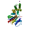
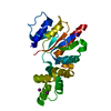
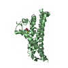
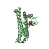
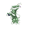
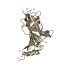
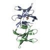
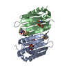
| Title | C-terminal region of the Xanthomonas campestris pv. campestris OLD protein phased with platinum | ||||||
 Components Components | Xcc_ctr_pt | ||||||
 Keywords Keywords | UNKNOWN FUNCTION / Nuclease / Toprim | ||||||
| Function / homology |  Function and homology information Function and homology information | ||||||
| Biological species |  Xanthomonas campestris pv. campestris (bacteria) Xanthomonas campestris pv. campestris (bacteria) | ||||||
| Method |  X-RAY DIFFRACTION / SYNCHROTRON / SAD / Resolution: 1.86 Å X-RAY DIFFRACTION / SYNCHROTRON / SAD / Resolution: 1.86 Å | ||||||
 Authors Authors | Schiltz, C.J. / Lee, A. / Partlow, E.A. / Hosford, C.J. / Chappie, J.S. | ||||||
 Citation Citation | Journal: Nucleic Acids Res. / Year: 2019 Title: Structural characterization of Class 2 OLD family nucleases supports a two-metal catalysis mechanism for cleavage. Authors: Schiltz, C.J. / Lee, A. / Partlow, E.A. / Hosford, C.J. / Chappie, J.S. | ||||||
| History |
|
- Structure visualization
| Structure viewer | Molecule: MolmilJmol/JSmol |
|---|
- Downloads & links
-Download
| PDBx/mmCIF format | 6njw.cif.gz | 67.6 KB | Display | PDBx/mmCIF format |
|---|---|---|---|---|
| PDB format | pdb6njw.ent.gz | 40 KB | Display | PDB format |
| PDBx/mmJSON format | 6njw.json.gz | Tree view | PDBx/mmJSON format | |
| Others |  Other downloads Other downloads |
-Validation report
| Arichive directory | https://data.pdbj.org/pub/pdb/validation_reports/nj/6njwftp://data.pdbj.org/pub/pdb/validation_reports/nj/6njw | HTTPS FTP |
|---|
-Related structure data


-Links
 PDBj
PDBj
- Assembly
| Deposited unit | 
| ||||||||||||
|---|---|---|---|---|---|---|---|---|---|---|---|---|---|
| 1 |
| ||||||||||||
| Unit cell |
|
-Components
| #1: Protein | Mass: 26036.361 Da / Num. of mol.: 1 Source method: isolated from a genetically manipulated source Source: (gene. exp.) Xanthomonas campestris pv. campestris (strain B100) (bacteria)Strain: B100 / Gene: XCCB100_2436 / Production host: | ||||
|---|---|---|---|---|---|
| #2: Chemical | ChemComp-PT /   Mass: 195.078 Da / Num. of mol.: 1 / Source method: obtained synthetically / Formula: Pt Mass: 195.078 Da / Num. of mol.: 1 / Source method: obtained synthetically / Formula: Pt | ||||
| #3: Chemical |   Mass: 126.904 Da / Num. of mol.: 2 / Source method: obtained synthetically / Formula: I Mass: 126.904 Da / Num. of mol.: 2 / Source method: obtained synthetically / Formula: I#4: Chemical | ChemComp-MG / |   Mass: 24.305 Da / Num. of mol.: 1 / Source method: obtained synthetically / Formula: Mg Mass: 24.305 Da / Num. of mol.: 1 / Source method: obtained synthetically / Formula: Mg#5: Water | ChemComp-HOH / |  Mass: 18.015 Da / Num. of mol.: 73 / Source method: isolated from a natural source / Formula: H2O Mass: 18.015 Da / Num. of mol.: 73 / Source method: isolated from a natural source / Formula: H2O |
-Experimental details
-Experiment
| Experiment | Method: X-RAY DIFFRACTION / Number of used crystals: 1 |
|---|
- Sample preparation
| Crystal | Density Matthews: 2.52 Å3/Da / Density % sol: 51.26 % |
|---|---|
| Crystal grow | Temperature: 293 K / Method: vapor diffusion, sitting drop Details: 0.1 M Bis Tris Propane pH 7.0-8.0, 11-26% PEG 3350, and 0.15-0.2 M sodium iodide |
-Data collection
| Diffraction | Mean temperature: 100 K / Serial crystal experiment: N |
|---|---|
| Diffraction source | Source: SYNCHROTRON / Site: APS  / Beamline: 24-ID-C / Wavelength: 1.0716 Å / Beamline: 24-ID-C / Wavelength: 1.0716 Å |
| Detector | Type: DECTRIS PILATUS 6M-F / Detector: PIXEL / Date: Dec 2, 2015 |
| Radiation | Protocol: SINGLE WAVELENGTH / Monochromatic (M) / Laue (L): M / Scattering type: x-ray |
| Radiation wavelength | Wavelength: 1.0716 Å / Relative weight: 1 |
| Reflection | Resolution: 1.86→65.43 Å / Num. obs: 44393 / % possible obs: 99.9 % / Redundancy: 10.2 % / Biso Wilson estimate: 42.91 Å2 / Net I/σ(I): 19.5 |
| Reflection shell | Resolution: 1.86→1.9 Å |
- Processing
| Software |
| |||||||||||||||||||||||||||||||||||||||||||||||||||||||||||||||||||||||||||||||||||||||||||||||||||||||||||||||||||||||
|---|---|---|---|---|---|---|---|---|---|---|---|---|---|---|---|---|---|---|---|---|---|---|---|---|---|---|---|---|---|---|---|---|---|---|---|---|---|---|---|---|---|---|---|---|---|---|---|---|---|---|---|---|---|---|---|---|---|---|---|---|---|---|---|---|---|---|---|---|---|---|---|---|---|---|---|---|---|---|---|---|---|---|---|---|---|---|---|---|---|---|---|---|---|---|---|---|---|---|---|---|---|---|---|---|---|---|---|---|---|---|---|---|---|---|---|---|---|---|---|---|
| Refinement | Method to determine structure: SAD / Resolution: 1.86→46.26 Å / SU ML: 0.287 / Cross valid method: FREE R-VALUE / σ(F): 1.77 / Phase error: 26.3421
| |||||||||||||||||||||||||||||||||||||||||||||||||||||||||||||||||||||||||||||||||||||||||||||||||||||||||||||||||||||||
| Solvent computation | Shrinkage radii: 0.9 Å / VDW probe radii: 1.11 Å | |||||||||||||||||||||||||||||||||||||||||||||||||||||||||||||||||||||||||||||||||||||||||||||||||||||||||||||||||||||||
| Displacement parameters | Biso mean: 49.24 Å2 | |||||||||||||||||||||||||||||||||||||||||||||||||||||||||||||||||||||||||||||||||||||||||||||||||||||||||||||||||||||||
| Refinement step | Cycle: LAST / Resolution: 1.86→46.26 Å
| |||||||||||||||||||||||||||||||||||||||||||||||||||||||||||||||||||||||||||||||||||||||||||||||||||||||||||||||||||||||
| Refine LS restraints |
| |||||||||||||||||||||||||||||||||||||||||||||||||||||||||||||||||||||||||||||||||||||||||||||||||||||||||||||||||||||||
| LS refinement shell |
|
