 Movie
Movie Controller


+ Open data

- Basic information
| Entry | Database: PDB / ID: 6neo | ||||||
|---|---|---|---|---|---|---|---|
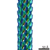
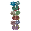
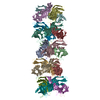
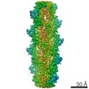
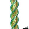
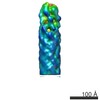
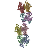
| Title | Crystal structure of PprA filament from Deinococcus radiodurans | ||||||
 Components Components | DNA repair protein PprA | ||||||
 Keywords Keywords | DNA BINDING PROTEIN / PROTEIN FIBRIL / DNA damage repair / Radiation induced / Genome segregation / Filament formation | ||||||
| Function / homology | cellular response to desiccation / cellular response to gamma radiation / double-strand break repair via nonhomologous end joining / double-stranded DNA binding / damaged DNA binding / DNA repair / DNA repair protein PprA Function and homology information Function and homology information | ||||||
| Biological species |  Deinococcus radiodurans (radioresistant) Deinococcus radiodurans (radioresistant) | ||||||
| Method |  X-RAY DIFFRACTION / MOLECULAR REPLACEMENT / Resolution: 5.94 Å X-RAY DIFFRACTION / MOLECULAR REPLACEMENT / Resolution: 5.94 Å | ||||||
 Authors Authors | Szabla, R. / Junop, M.S. / Rok, M. | ||||||
| Funding support |  Canada, 1items Canada, 1items
| ||||||
 Citation Citation | Journal: To Be Published Title: Crystal structure of PprA filament from Deinococcus radiodurans Authors: Szabla, R. / Junop, M.S. / Rok, M. | ||||||
| History |
|
- Structure visualization
| Structure viewer | Molecule: MolmilJmol/JSmol |
|---|
- Downloads & links
-Download
| PDBx/mmCIF format | 6neo.cif.gz | 57.5 KB | Display | PDBx/mmCIF format |
|---|---|---|---|---|
| PDB format | pdb6neo.ent.gz | 36.6 KB | Display | PDB format |
| PDBx/mmJSON format | 6neo.json.gz | Tree view | PDBx/mmJSON format | |
| Others |  Other downloads Other downloads |
-Validation report
| Summary document | 6neo_validation.pdf.gz | 387.8 KB | Display | wwPDB validaton report |
|---|---|---|---|---|
| Full document | 6neo_full_validation.pdf.gz | 387.8 KB | Display | |
| Data in XML | 6neo_validation.xml.gz | 6.4 KB | Display | |
| Data in CIF | 6neo_validation.cif.gz | 8.6 KB | Display | |
| Arichive directory | https://data.pdbj.org/pub/pdb/validation_reports/ne/6neoftp://data.pdbj.org/pub/pdb/validation_reports/ne/6neo | HTTPS FTP |
-Related structure data
| Related structure data |  6mc8S S: Starting model for refinement |
|---|---|
| Similar structure data |

-Links
 PDBj
PDBj- Assembly
| Deposited unit | 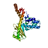
| ||||||||
|---|---|---|---|---|---|---|---|---|---|
| 1 | 
| ||||||||
| Unit cell |
|
-Components
| #1: Protein | Mass: 29919.477 Da / Num. of mol.: 1 / Mutation: D180K, D184K Source method: isolated from a genetically manipulated source Source: (gene. exp.) Deinococcus radiodurans (strain ATCC 13939 / DSM 20539 / JCM 16871 / LMG 4051 / NBRC 15346 / NCIMB 9279 / R1 / VKM B-1422) (radioresistant)Strain: ATCC 13939 / DSM 20539 / JCM 16871 / LMG 4051 / NBRC 15346 / NCIMB 9279 / R1 / VKM B-1422 Gene: pprA, DR_A0346 / Plasmid: pDEST-527 Details (production host): Gateway destination vector with TEV-protease cleavable N-terminal His tag Production host: |
|---|
-Experimental details
-Experiment
| Experiment | Method: X-RAY DIFFRACTION / Number of used crystals: 1 |
|---|
- Sample preparation
| Crystal | Density Matthews: 4.55 Å3/Da / Density % sol: 72.96 % / Description: long hexagonal-base prism |
|---|---|
| Crystal grow | Temperature: 293.15 K / Method: vapor diffusion, hanging drop / pH: 6.5 Details: 1:1 4.9 mg/mL protein in 150 mM potassium chloride, 20 mM Tris, pH 8.0 + Morpheus II-FX96 (Molecular Dimensions) condition H2 - 40 mM polyamines (spermine tetrahydrochloride, spermidine ...Details: 1:1 4.9 mg/mL protein in 150 mM potassium chloride, 20 mM Tris, pH 8.0 + Morpheus II-FX96 (Molecular Dimensions) condition H2 - 40 mM polyamines (spermine tetrahydrochloride, spermidine trihydrochloride, 1,4-diaminobutane dihydrochloride, DL-ornithine monohydrochloride), 50% v/v Precipitant Mix 6 (25% w/v PEG4000, 40% w/v 1,2,6-hexanetriol), 0.1 M Buffer System 4 (Gly-Gly, AMPD) at pH 6.5. The drop was suspended over a 1.5 M ammonium sulfate dehydrating solution and incubated at 20 degrees C for about 5 months. Temp details: Temperature-controlled incubator |
-Data collection
| Diffraction | Mean temperature: 100 K / Ambient temp details: Nitrogen gas cryostream / Serial crystal experiment: N |
|---|---|
| Diffraction source | Source: ROTATING ANODE / Type: RIGAKU MICROMAX-007 HF / Wavelength: 1.5418 Å |
| Detector | Type: RIGAKU SATURN 944+ / Detector: CCD / Date: Jun 8, 2018 |
| Radiation | Protocol: SINGLE WAVELENGTH / Monochromatic (M) / Laue (L): M / Scattering type: x-ray |
| Radiation wavelength | Wavelength: 1.5418 Å / Relative weight: 1 |
| Reflection | Resolution: 5.94→67.53 Å / Num. obs: 1513 / % possible obs: 98.8 % / Redundancy: 13.9 % / CC1/2: 0.995 / Rmerge(I) obs: 0.261 / Rpim(I) all: 0.074 / Rrim(I) all: 0.278 / Χ2: 0.97 / Net I/σ(I): 9.5 |
| Reflection shell | Resolution: 5.94→6.64 Å / Redundancy: 13.8 % / Mean I/σ(I) obs: 1.4 / Num. unique obs: 402 / CC1/2: 0.496 / Rpim(I) all: 0.565 / Χ2: 0.92 / % possible all: 96.8 |
- Processing
| Software |
| ||||||||||||||||||||||||
|---|---|---|---|---|---|---|---|---|---|---|---|---|---|---|---|---|---|---|---|---|---|---|---|---|---|
| Refinement | Method to determine structure: MOLECULAR REPLACEMENT Starting model: PDB entry 6MC8 chain B Resolution: 5.94→50.0319 Å / SU ML: 0.41 / Cross valid method: FREE R-VALUE / σ(F): 1.34 / Phase error: 27.03
| ||||||||||||||||||||||||
| Solvent computation | Shrinkage radii: 0.9 Å / VDW probe radii: 1.11 Å | ||||||||||||||||||||||||
| Refinement step | Cycle: LAST / Resolution: 5.94→50.0319 Å
| ||||||||||||||||||||||||
| Refine LS restraints |
| ||||||||||||||||||||||||
| LS refinement shell | Highest resolution: 5.9395 Å |
