 Movie
Movie Controller


+ Open data

- Basic information
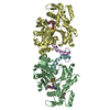
| Entry | Database: PDB / ID: 6mc8 | ||||||
|---|---|---|---|---|---|---|---|
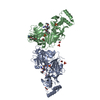
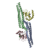
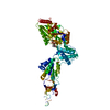
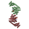
| Title | Crystal structure of PprA dimer from Deinococcus deserti | ||||||
 Components Components | DNA repair protein PprA | ||||||
 Keywords Keywords | DNA BINDING PROTEIN / DNA damage repair / Radiation induced / Genome segregation / Filament formation | ||||||
| Function / homology | Aspartyl protease, retroviral-type family profile. / Peptidase A2A, retrovirus, catalytic / aspartic-type endopeptidase activity / proteolysis / Putative DNA repair protein PprA Function and homology information Function and homology information | ||||||
| Biological species |  Deinococcus deserti (bacteria) Deinococcus deserti (bacteria) | ||||||
| Method |  X-RAY DIFFRACTION / MOLECULAR REPLACEMENT / Resolution: 2.5 Å X-RAY DIFFRACTION / MOLECULAR REPLACEMENT / Resolution: 2.5 Å | ||||||
 Authors Authors | Szabla, R. / Junop, M.S. / Rok, M. | ||||||
| Funding support |  Canada, 1items Canada, 1items
| ||||||
 Citation Citation | Journal: To Be Published Title: Crystal structure of PprA from Deinococcus radiodurans Authors: Szabla, R. / Czerwinski, M. / Junop, M.S. | ||||||
| History |
|
- Structure visualization
| Structure viewer | Molecule: MolmilJmol/JSmol |
|---|
- Downloads & links
-Download
| PDBx/mmCIF format | 6mc8.cif.gz | 120.6 KB | Display | PDBx/mmCIF format |
|---|---|---|---|---|
| PDB format | pdb6mc8.ent.gz | 90.9 KB | Display | PDB format |
| PDBx/mmJSON format | 6mc8.json.gz | Tree view | PDBx/mmJSON format | |
| Others |  Other downloads Other downloads |
-Validation report
| Arichive directory | https://data.pdbj.org/pub/pdb/validation_reports/mc/6mc8ftp://data.pdbj.org/pub/pdb/validation_reports/mc/6mc8 | HTTPS FTP |
|---|
-Related structure data


-Links
 PDBj
PDBj- Assembly
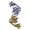
| Deposited unit | 
| ||||||||||||
|---|---|---|---|---|---|---|---|---|---|---|---|---|---|
| 1 |
| ||||||||||||
| Unit cell |
|
-Components
| #1: Protein | Mass: 33631.777 Da / Num. of mol.: 2 / Mutation: D192K, D196K Source method: isolated from a genetically manipulated source Source: (gene. exp.) Deinococcus deserti (strain VCD115 / DSM 17065 / LMG 22923) (bacteria)Strain: VCD115 / DSM 17065 / LMG 22923 / Gene: pprA, Deide_2p01380 / Plasmid: pDEST-527 Details (production host): Gateway destination vector for bacterial expression; His tag Production host: #2: Water | ChemComp-HOH / |  Mass: 18.015 Da / Num. of mol.: 174 / Source method: isolated from a natural source / Formula: H2O Mass: 18.015 Da / Num. of mol.: 174 / Source method: isolated from a natural source / Formula: H2O |
|---|
-Experimental details
-Experiment
| Experiment | Method: X-RAY DIFFRACTION / Number of used crystals: 1 |
|---|
- Sample preparation
| Crystal | Density Matthews: 2.59 Å3/Da / Density % sol: 52.51 % / Description: square bipyramid |
|---|---|
| Crystal grow | Temperature: 293.15 K / Method: vapor diffusion, hanging drop / pH: 9.5 Details: Protein at 3.0 mg/mL in 800 mM NaCl, 150 mM Imidazole, 20 mM Tris, pH 8.0 was mixed in 1:1 volume ratio with MCSG3 #92 - a solution of 30 % (w/v) PEG 400 and 0.1 M CHES, pH 9.5. The drop was ...Details: Protein at 3.0 mg/mL in 800 mM NaCl, 150 mM Imidazole, 20 mM Tris, pH 8.0 was mixed in 1:1 volume ratio with MCSG3 #92 - a solution of 30 % (w/v) PEG 400 and 0.1 M CHES, pH 9.5. The drop was suspended over 1.5M Ammonium sulfate. Temp details: Constant-temperature incubation |
-Data collection
| Diffraction | Mean temperature: 100 K / Ambient temp details: Nitrogen gas cryostream |
|---|---|
| Diffraction source | Source: ROTATING ANODE / Type: RIGAKU MICROMAX-007 HF / Wavelength: 1.54056 Å |
| Detector | Type: RIGAKU SATURN 944+ / Detector: CCD / Date: Jun 30, 2017 |
| Radiation | Protocol: SINGLE WAVELENGTH / Monochromatic (M) / Laue (L): M / Scattering type: x-ray |
| Radiation wavelength | Wavelength: 1.54056 Å / Relative weight: 1 |
| Reflection | Resolution: 2.5→28.72 Å / Num. obs: 22737 / % possible obs: 96.7 % / Redundancy: 24.1 % / CC1/2: 0.998 / Rmerge(I) obs: 0.182 / Rpim(I) all: 0.049 / Rrim(I) all: 0.189 / Net I/σ(I): 19.8 |
| Reflection shell | Resolution: 2.5→2.6 Å / Redundancy: 11.6 % / Rmerge(I) obs: 0.663 / Mean I/σ(I) obs: 2 / Num. unique obs: 2079 / CC1/2: 0.845 / Rpim(I) all: 0.223 / Rrim(I) all: 0.7 / % possible all: 80.2 |
- Processing
| Software |
| ||||||||||||||||||
|---|---|---|---|---|---|---|---|---|---|---|---|---|---|---|---|---|---|---|---|
| Refinement | Method to determine structure: MOLECULAR REPLACEMENT / Resolution: 2.5→28.72 Å / Cross valid method: FREE R-VALUE
| ||||||||||||||||||
| Refinement step | Cycle: LAST / Resolution: 2.5→28.72 Å
|
