- PDB-6mc6: Crystal structure of PprA filament from Deinococcus radiodurans -
+
Open data
ID or keywords:
Loading...
-
Basic information
Entry
Database: PDB / ID: 6mc6
Title
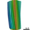
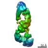
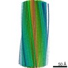
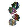
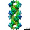
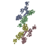
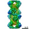
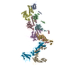
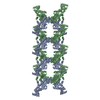
Crystal structure of PprA filament from Deinococcus radiodurans
Components
DNA repair protein PprA
Keywords
DNA BINDING PROTEIN / DNA damage repair / Radiation induced / Genome segregation / Filment formation
Function / homology
cellular response to desiccation / cellular response to gamma radiation / double-strand break repair via nonhomologous end joining / double-stranded DNA binding / damaged DNA binding / DNA repair / DNA repair protein PprA
A: DNA repair protein PprA B: DNA repair protein PprA
A: DNA repair protein PprA B: DNA repair protein PprA
A: DNA repair protein PprA B: DNA repair protein PprA
A: DNA repair protein PprA B: DNA repair protein PprA
A: DNA repair protein PprA B: DNA repair protein PprA
A: DNA repair protein PprA B: DNA repair protein PprA
A: DNA repair protein PprA B: DNA repair protein PprA
A: DNA repair protein PprA B: DNA repair protein PprA
defined by author
Evidence: gel filtration, Purified PprA elutes from a gel filtration column at a broad distribution of retention volumes. The retention volume corresponds to an oligomeric size ranging from a ...Evidence: gel filtration, Purified PprA elutes from a gel filtration column at a broad distribution of retention volumes. The retention volume corresponds to an oligomeric size ranging from a tetramer to a 50-mer and larger., scanning transmission electron microscopy, In vitro, PprA exists as a long filament of varying length, as observed by TEM. This behavior is independent of Deinococcus species of origin., microscopy, In vivo, PprA forms a thin strand spanning the septum of dividing D.radiodurans cells. This was visualized by fluorescence microscopy., The repeating filament assembly present in this crystal has been observed in numerous other crystal forms, despite different symmetries and crystal packing., Residues that are required for DNA binding all map to two distinct faces of this assembly.
Mass: 18.015 Da / Num. of mol.: 44 / Source method: isolated from a natural source / Formula: H2O
-
Experimental details
-
Experiment
Experiment
Method: X-RAY DIFFRACTION / Number of used crystals: 1
-
Sample preparation
Crystal
Density Matthews: 2.5 Å3/Da / Density % sol: 50.8 % / Description: long rectangular prism
Crystal grow
Temperature: 293.15 K / Method: vapor diffusion, hanging drop / pH: 8 Details: Protein at 4.5 mg/mL in 150 mM KCl, 20 mM Tris, pH 7.5 was mixed in 1:1 volume ratio with Wizard Classic 1 (Rigaku) #46 - a solution of 200 mM Lithium sulfate, 10% (w/v) PEG 8000 and 100 mM ...Details: Protein at 4.5 mg/mL in 150 mM KCl, 20 mM Tris, pH 7.5 was mixed in 1:1 volume ratio with Wizard Classic 1 (Rigaku) #46 - a solution of 200 mM Lithium sulfate, 10% (w/v) PEG 8000 and 100 mM Imidazole/ HCl at pH 8.0. The drop was suspended over a 1.5M Ammonium sulfate dehydrating solution and incubated for about 5 weeks. Temp details: Temperature-controlled incubation
-
Data collection
Diffraction
Mean temperature: 100 K / Ambient temp details: Nitrogen gas cryo-stream
In the structure databanks used in Yorodumi, some data are registered as the other names, "COVID-19 virus" and "2019-nCoV". Here are the details of the virus and the list of structure data.
Jan 31, 2019. EMDB accession codes are about to change! (news from PDBe EMDB page)
EMDB accession codes are about to change! (news from PDBe EMDB page)
The allocation of 4 digits for EMDB accession codes will soon come to an end. Whilst these codes will remain in use, new EMDB accession codes will include an additional digit and will expand incrementally as the available range of codes is exhausted. The current 4-digit format prefixed with “EMD-” (i.e. EMD-XXXX) will advance to a 5-digit format (i.e. EMD-XXXXX), and so on. It is currently estimated that the 4-digit codes will be depleted around Spring 2019, at which point the 5-digit format will come into force.
The EM Navigator/Yorodumi systems omit the EMD- prefix.
Related info.:Q: What is EMD? / ID/Accession-code notation in Yorodumi/EM Navigator
Yorodumi is a browser for structure data from EMDB, PDB, SASBDB, etc.
This page is also the successor to EM Navigator detail page, and also detail information page/front-end page for Omokage search.
The word "yorodu" (or yorozu) is an old Japanese word meaning "ten thousand". "mi" (miru) is to see.
Related info.:EMDB / PDB / SASBDB / Comparison of 3 databanks / Yorodumi Search / Aug 31, 2016. New EM Navigator & Yorodumi / Yorodumi Papers / Jmol/JSmol / Function and homology information / Changes in new EM Navigator and Yorodumi
 Movie
Movie Controller


 Open data
 Basic information
 Components
 Keywords
 Function and homology information
 Deinococcus radiodurans R1 (radioresistant)
 X-RAY DIFFRACTION /
  Authors
 Canada, 1items
  Citation
 Structure visualization
 Downloads & links
 Other downloads

 PDBj
PDBj Assembly
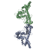
 Mass: 18.015 Da / Num. of mol.: 44 / Source method: isolated from a natural source / Formula: H2O
 Sample preparation
 / Beamline: 17-ID / Wavelength: 0.97928 Å
 Processing