 Movie
Movie Controller


+ Open data

- Basic information
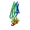
| Entry | Database: PDB / ID: 4u0z | ||||||
|---|---|---|---|---|---|---|---|
| Title | Eukaryotic Fic Domain containing protein with bound APCPP | ||||||
 Components Components | Adenosine monophosphate-protein transferase FICD | ||||||
 Keywords Keywords | TRANSFERASE / TPR / FIC / APCPP / adenylation | ||||||
| Function / homology |  Function and homology information Function and homology informationprotein deadenylylation / protein adenylylhydrolase activity / protein adenylylation / AMPylase activity / protein adenylyltransferase / regulation of IRE1-mediated unfolded protein response / negative regulation of GTPase activity / Hydrolases; Acting on ester bonds; Phosphoric-diester hydrolases / response to unfolded protein / Hsp70 protein binding ...protein deadenylylation / protein adenylylhydrolase activity / protein adenylylation / AMPylase activity / protein adenylyltransferase / regulation of IRE1-mediated unfolded protein response / negative regulation of GTPase activity / Hydrolases; Acting on ester bonds; Phosphoric-diester hydrolases / response to unfolded protein / Hsp70 protein binding / response to endoplasmic reticulum stress / protein-folding chaperone binding / endoplasmic reticulum membrane / endoplasmic reticulum / protein homodimerization activity / ATP binding / identical protein binding Similarity search - Function | ||||||
| Biological species |  Homo sapiens (human) Homo sapiens (human) | ||||||
| Method |  X-RAY DIFFRACTION / SYNCHROTRON / MOLECULAR REPLACEMENT / Resolution: 2.95 Å X-RAY DIFFRACTION / SYNCHROTRON / MOLECULAR REPLACEMENT / Resolution: 2.95 Å | ||||||
 Authors Authors | Cole, A.R. / Bunney, T.D. / Katan, M. | ||||||
 Citation Citation | Journal: Structure / Year: 2014 Title: Crystal structure of the human, FIC-domain containing protein HYPE and implications for its functions. Authors: Bunney, T.D. / Cole, A.R. / Broncel, M. / Esposito, D. / Tate, E.W. / Katan, M. | ||||||
| History |
|
- Structure visualization
| Structure viewer | Molecule: MolmilJmol/JSmol |
|---|
- Downloads & links
-Download
| PDBx/mmCIF format | 4u0z.cif.gz | 531.3 KB | Display | PDBx/mmCIF format |
|---|---|---|---|---|
| PDB format | pdb4u0z.ent.gz | 434.3 KB | Display | PDB format |
| PDBx/mmJSON format | 4u0z.json.gz | Tree view | PDBx/mmJSON format | |
| Others |  Other downloads Other downloads |
-Validation report
| Arichive directory | https://data.pdbj.org/pub/pdb/validation_reports/u0/4u0zftp://data.pdbj.org/pub/pdb/validation_reports/u0/4u0z | HTTPS FTP |
|---|
-Related structure data
| Related structure data |  4u04C  4u07C  4u0sC  4u0uC  3cucS S: Starting model for refinement C: citing same article ( |
|---|---|
| Similar structure data |


-Links
 PDBj
PDBj


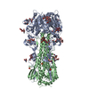
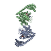
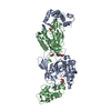
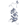
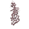
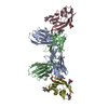
- Assembly
| Deposited unit | 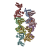
| ||||||||
|---|---|---|---|---|---|---|---|---|---|
| 1 | 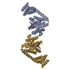
| ||||||||
| 2 | 
| ||||||||
| 3 | 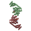
| ||||||||
| 4 | 
| ||||||||
| 5 | 
| ||||||||
| 6 | 
| ||||||||
| 7 | 
| ||||||||
| Unit cell |
|
-Components
| #1: Protein | Mass: 39548.273 Da / Num. of mol.: 8 / Fragment: UNP residues 102-445 Source method: isolated from a genetically manipulated source Source: (gene. exp.) Homo sapiens (human) / Gene: FICD, HIP13, HYPE, UNQ3041/PRO9857 / Production host:  References: UniProt: Q9BVA6, Transferases; Transferring phosphorus-containing groups; Nucleotidyltransferases #2: Chemical | ChemComp-APC /   Mass: 505.208 Da / Num. of mol.: 8 / Source method: obtained synthetically / Formula: C11H18N5O12P3 / Comment: AMP-CPP, energy-carrying molecule analogue*YM Mass: 505.208 Da / Num. of mol.: 8 / Source method: obtained synthetically / Formula: C11H18N5O12P3 / Comment: AMP-CPP, energy-carrying molecule analogue*YM#3: Chemical | ChemComp-MG /   Mass: 24.305 Da / Num. of mol.: 8 / Source method: obtained synthetically / Formula: Mg Mass: 24.305 Da / Num. of mol.: 8 / Source method: obtained synthetically / Formula: Mg#4: Water | ChemComp-HOH / |  Mass: 18.015 Da / Num. of mol.: 816 / Source method: isolated from a natural source / Formula: H2O Mass: 18.015 Da / Num. of mol.: 816 / Source method: isolated from a natural source / Formula: H2O |
|---|
-Experimental details
-Experiment
| Experiment | Method: X-RAY DIFFRACTION |
|---|
- Sample preparation
| Crystal | Density Matthews: 2.65 Å3/Da / Density % sol: 53.65 % |
|---|---|
| Crystal grow | Temperature: 292 K / Method: vapor diffusion, sitting drop / pH: 7.5 Details: 25% PEG 3350, 200mM Na K Tartrate, 100mM Bis-Tris Propane 7.5 |
-Data collection
| Diffraction | Mean temperature: 100 K |
|---|---|
| Diffraction source | Source: SYNCHROTRON / Site: Diamond  / Beamline: I03 / Wavelength: 0.9763 Å / Beamline: I03 / Wavelength: 0.9763 Å |
| Detector | Type: PSI PILATUS 6M / Detector: PIXEL / Date: Sep 26, 2012 |
| Radiation | Protocol: SINGLE WAVELENGTH / Monochromatic (M) / Laue (L): M / Scattering type: x-ray |
| Radiation wavelength | Wavelength: 0.9763 Å / Relative weight: 1 |
| Reflection | Resolution: 2.95→43.34 Å / Num. obs: 65073 / % possible obs: 94.7 % / Redundancy: 3.5 % / Biso Wilson estimate: 55.82 Å2 / Net I/σ(I): 4.4 |
| Reflection shell | Resolution: 2.95→3.02 Å / Redundancy: 3.1 % / Rmerge(I) obs: 0.496 / Mean I/σ(I) obs: 1.7 |
- Processing
| Software |
| ||||||||||||||||||||||||||||||||||||||||||||||||||||||||||||||||||||||||||||||||||||||||||||||||||||||||||||||||||
|---|---|---|---|---|---|---|---|---|---|---|---|---|---|---|---|---|---|---|---|---|---|---|---|---|---|---|---|---|---|---|---|---|---|---|---|---|---|---|---|---|---|---|---|---|---|---|---|---|---|---|---|---|---|---|---|---|---|---|---|---|---|---|---|---|---|---|---|---|---|---|---|---|---|---|---|---|---|---|---|---|---|---|---|---|---|---|---|---|---|---|---|---|---|---|---|---|---|---|---|---|---|---|---|---|---|---|---|---|---|---|---|---|---|---|---|
| Refinement | Method to determine structure: MOLECULAR REPLACEMENT Starting model: 3cuc Resolution: 2.95→43.34 Å / Cor.coef. Fo:Fc: 0.8842 / Cor.coef. Fo:Fc free: 0.8616 / Cross valid method: THROUGHOUT / σ(F): 0 / SU Rfree Blow DPI: 0.418
| ||||||||||||||||||||||||||||||||||||||||||||||||||||||||||||||||||||||||||||||||||||||||||||||||||||||||||||||||||
| Displacement parameters | Biso mean: 61.13 Å2
| ||||||||||||||||||||||||||||||||||||||||||||||||||||||||||||||||||||||||||||||||||||||||||||||||||||||||||||||||||
| Refine analyze | Luzzati coordinate error obs: 0.4 Å | ||||||||||||||||||||||||||||||||||||||||||||||||||||||||||||||||||||||||||||||||||||||||||||||||||||||||||||||||||
| Refinement step | Cycle: 1 / Resolution: 2.95→43.34 Å
| ||||||||||||||||||||||||||||||||||||||||||||||||||||||||||||||||||||||||||||||||||||||||||||||||||||||||||||||||||
| Refine LS restraints |
| ||||||||||||||||||||||||||||||||||||||||||||||||||||||||||||||||||||||||||||||||||||||||||||||||||||||||||||||||||
| LS refinement shell | Resolution: 2.95→3.03 Å / Total num. of bins used: 20
|
