 Movie
Movie Controller


+ Open data

- Basic information
| Entry | Database: EMDB / ID: EMD-1240 | |||||||||
|---|---|---|---|---|---|---|---|---|---|---|
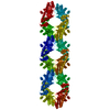
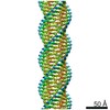
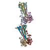
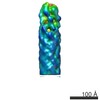
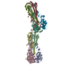
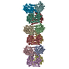
| Title | The structure of a filamentous bacteriophage. | |||||||||
 Map data Map data | This is the volume generated by IHRSR. To align with PDB 2hi5, use "CP TO BRIX" in SPIDER, with 2.4 A/pixel, and default unit cell. | |||||||||
 Sample Sample |
| |||||||||
| Function / homology | Phage major coat protein, Gp8 / Bacteriophage M13, G8P, capsid domain superfamily / Capsid protein G8P / helical viral capsid / host cell membrane / Capsid protein G8P Function and homology information Function and homology information | |||||||||
| Biological species |  Enterobacteria phage fd (virus) Enterobacteria phage fd (virus) | |||||||||
| Method | helical reconstruction / cryo EM / negative staining / Resolution: 8.0 Å | |||||||||
 Authors Authors | Wang YA / Yu X / Overman S / Tsuboi M / Thomas GJ / Egelman EH | |||||||||
 Citation Citation | Journal: J Mol Biol / Year: 2006 Title: The structure of a filamentous bacteriophage. Authors: Ying A Wang / Xiong Yu / Stacy Overman / Masamichi Tsuboi / George J Thomas / Edward H Egelman /  Abstract: Many thin helical polymers, including bacterial pili and filamentous bacteriophage, have been seen as refractory to high-resolution studies by electron microscopy. Studies of the quaternary structure ...Many thin helical polymers, including bacterial pili and filamentous bacteriophage, have been seen as refractory to high-resolution studies by electron microscopy. Studies of the quaternary structure of such filaments have depended upon techniques such as modeling or X-ray fiber diffraction, given that direct visualization of the subunit organization has not been possible. We report the first image reconstruction of a filamentous virus, bacteriophage fd, by cryoelectron microscopy. Although these thin ( approximately 70 A in diameter) rather featureless filaments scatter weakly, we have been able to achieve a nominal resolution of approximately 8 A using an iterative helical reconstruction procedure. We show that two different conformations of the virus exist, and that in both states the subunits are packed differently than in conflicting models previously proposed on the basis of X-ray fiber diffraction or solid-state NMR studies. A significant fraction of the population of wild-type fd is either disordered or in multiple conformational states, while in the presence of the Y21M mutation, this heterogeneity is greatly reduced, consistent with previous observations. These results show that new computational approaches to helical reconstruction can greatly extend the ability to visualize heterogeneous protein polymers at a reasonably high resolution. | |||||||||
| History |
|
- Structure visualization
| Movie |
Movie viewer |
|---|---|
| Structure viewer | EM map: SurfViewMolmilJmol/JSmol |
| Supplemental images |

- Downloads & links
-EMDB archive
| Map data | emd_1240.map.gz | 368.1 KB | EMDB map data format | |
|---|---|---|---|---|
| Header (meta data) | emd-1240-v30.xmlemd-1240.xml | 9.1 KB 9.1 KB | Display Display | EMDB header |
| Images |  1240.gif 1240.gif | 9.1 KB | ||
| Archive directory |  http://ftp.pdbj.org/pub/emdb/structures/EMD-1240ftp://ftp.pdbj.org/pub/emdb/structures/EMD-1240 http://ftp.pdbj.org/pub/emdb/structures/EMD-1240ftp://ftp.pdbj.org/pub/emdb/structures/EMD-1240 | HTTPS FTP |
-Related structure data
| Related structure data | 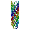 2hi5MC M: atomic model generated by this map C: citing same article ( |
|---|---|
| Similar structure data |


-Links
| EMDB pages | EMDB (EBI/PDBe) / EMDataResource |
|---|
-Map
| File | Download / File: emd_1240.map.gz / Format: CCP4 / Size: 3.7 MB / Type: IMAGE STORED AS FLOATING POINT NUMBER (4 BYTES) | ||||||||||||||||||||||||||||||||||||||||||||||||||||||||||||
|---|---|---|---|---|---|---|---|---|---|---|---|---|---|---|---|---|---|---|---|---|---|---|---|---|---|---|---|---|---|---|---|---|---|---|---|---|---|---|---|---|---|---|---|---|---|---|---|---|---|---|---|---|---|---|---|---|---|---|---|---|---|
| Annotation | This is the volume generated by IHRSR. To align with PDB 2hi5, use "CP TO BRIX" in SPIDER, with 2.4 A/pixel, and default unit cell. | ||||||||||||||||||||||||||||||||||||||||||||||||||||||||||||
| Projections & slices | Image control
Images are generated by Spider. | ||||||||||||||||||||||||||||||||||||||||||||||||||||||||||||
| Voxel size | X=Y=Z: 2.4 Å | ||||||||||||||||||||||||||||||||||||||||||||||||||||||||||||
| Density |
| ||||||||||||||||||||||||||||||||||||||||||||||||||||||||||||
| Symmetry | Space group: 1 | ||||||||||||||||||||||||||||||||||||||||||||||||||||||||||||
| Details | EMDB XML:
CCP4 map header:
| ||||||||||||||||||||||||||||||||||||||||||||||||||||||||||||
 Z (Sec.)
 Y (Row.)
 X (Col.)


-Supplemental data
- Sample components
-Entire : bacteriophage fd
| Entire | Name: bacteriophage fd (virus) |
|---|---|
| Components |
|
-Supramolecule #1000: bacteriophage fd
| Supramolecule | Name: bacteriophage fd / type: sample / ID: 1000 / Oligomeric state: polymer of single protein / Number unique components: 1 |
|---|
-Macromolecule #1: pVIII
| Macromolecule | Name: pVIII / type: protein_or_peptide / ID: 1 / Recombinant expression: No / Database: NCBI |
|---|---|
| Source (natural) | Organism: Enterobacteria phage fd (virus) / synonym: bacteriophage fd |
| Molecular weight | Experimental: 5207 MDa |
-Experimental details
-Structure determination
| Method | negative staining, cryo EM |
|---|---|
 Processing Processing | helical reconstruction |
| Aggregation state | filament |
-Sample preparation
| Staining | Type: NEGATIVE / Details: unstained |
|---|---|
| Vitrification | Cryogen name: ETHANE / Method: Blot for 2 seconds before plunging |
- Electron microscopy
| Microscope | FEI TECNAI F20 |
|---|---|
| Image recording | Category: FILM / Film or detector model: KODAK SO-163 FILM / Digitization - Scanner: OTHER / Digitization - Sampling interval: 6.35 µm / Number real images: 67 / Bits/pixel: 14 |
| Electron beam | Acceleration voltage: 200 kV / Electron source:  FIELD EMISSION GUN FIELD EMISSION GUN |
| Electron optics | Illumination mode: FLOOD BEAM / Imaging mode: BRIGHT FIELD / Nominal defocus max: 3.8 µm / Nominal defocus min: 1.4 µm / Nominal magnification: 50000 |
| Sample stage | Specimen holder: 626 / Specimen holder model: GATAN LIQUID NITROGEN |
| Experimental equipment |  Model: Tecnai F20 / Image courtesy: FEI Company |
-Image processing
| Final reconstruction | Applied symmetry - Helical parameters - Δz: 17.4 Å Applied symmetry - Helical parameters - Δ&Phi: 37.4 ° Applied symmetry - Helical parameters - Axial symmetry: C5 (5 fold cyclic) Algorithm: OTHER / Resolution.type: BY AUTHOR / Resolution: 8.0 Å / Resolution method: FSC 0.5 CUT-OFF / Software - Name: IHRSR |
|---|---|
| CTF correction | Details: each film |
