 Movie
Movie Controller

[English] 日本語
 Yorodumi
- PDB-6n4f: The crystal structure of hemagglutinin from A/canine/IL/11613/201... -
+ Open data

- Basic information
| Entry | Database: PDB / ID: 6n4f | ||||||
|---|---|---|---|---|---|---|---|
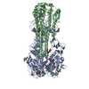
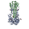
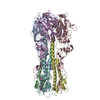
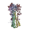
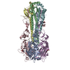
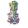
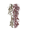
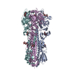
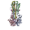
| Title | The crystal structure of hemagglutinin from A/canine/IL/11613/2015 (H3N2) influenza virus. | ||||||
 Components Components |
| ||||||
 Keywords Keywords | VIRAL PROTEIN / Hemagglutinin / canine influenza virus | ||||||
| Function / homology |  Function and homology information Function and homology informationviral budding from plasma membrane / clathrin-dependent endocytosis of virus by host cell / apical plasma membrane / host cell surface receptor binding / fusion of virus membrane with host plasma membrane / fusion of virus membrane with host endosome membrane / viral envelope / virion attachment to host cell / host cell plasma membrane / virion membrane Similarity search - Function | ||||||
| Biological species |   unidentified influenza virus unidentified influenza virus | ||||||
| Method |  X-RAY DIFFRACTION / SYNCHROTRON / MOLECULAR REPLACEMENT / Resolution: 3.01 Å X-RAY DIFFRACTION / SYNCHROTRON / MOLECULAR REPLACEMENT / Resolution: 3.01 Å | ||||||
 Authors Authors | Yang, H. / Stevens, J. | ||||||
 Citation Citation | Journal: J. Infect. Dis. / Year: 2017 Title: Assessment of Molecular, Antigenic, and Pathological Features of Canine Influenza A(H3N2) Viruses That Emerged in the United States. Authors: Pulit-Penaloza, J.A. / Simpson, N. / Yang, H. / Creager, H.M. / Jones, J. / Carney, P. / Belser, J.A. / Yang, G. / Chang, J. / Zeng, H. / Thor, S. / Jang, Y. / Killian, M.L. / Jenkins-Moore, ...Authors: Pulit-Penaloza, J.A. / Simpson, N. / Yang, H. / Creager, H.M. / Jones, J. / Carney, P. / Belser, J.A. / Yang, G. / Chang, J. / Zeng, H. / Thor, S. / Jang, Y. / Killian, M.L. / Jenkins-Moore, M. / Janas-Martindale, A. / Dubovi, E. / Wentworth, D.E. / Stevens, J. / Tumpey, T.M. / Davis, C.T. / Maines, T.R. | ||||||
| History |
|
- Structure visualization
| Structure viewer | Molecule: MolmilJmol/JSmol |
|---|
- Downloads & links
-Download
| PDBx/mmCIF format | 6n4f.cif.gz | 386.2 KB | Display | PDBx/mmCIF format |
|---|---|---|---|---|
| PDB format | pdb6n4f.ent.gz | 314.5 KB | Display | PDB format |
| PDBx/mmJSON format | 6n4f.json.gz | Tree view | PDBx/mmJSON format | |
| Others |  Other downloads Other downloads |
-Validation report
| Arichive directory | https://data.pdbj.org/pub/pdb/validation_reports/n4/6n4fftp://data.pdbj.org/pub/pdb/validation_reports/n4/6n4f | HTTPS FTP |
|---|
-Related structure data


-Links
 PDBj
PDBj


- Assembly
| Deposited unit | 
| |||||||||||||||||||||||||||||||||||||||||||||||||||||||||||||||||||||||||||||||||||||||||||||||||||||||||||||||||||||||||||||||||||||||||||||||||||||||||||||||||||||||||||||||||||||||||||||||||||||||||||||||||||||
|---|---|---|---|---|---|---|---|---|---|---|---|---|---|---|---|---|---|---|---|---|---|---|---|---|---|---|---|---|---|---|---|---|---|---|---|---|---|---|---|---|---|---|---|---|---|---|---|---|---|---|---|---|---|---|---|---|---|---|---|---|---|---|---|---|---|---|---|---|---|---|---|---|---|---|---|---|---|---|---|---|---|---|---|---|---|---|---|---|---|---|---|---|---|---|---|---|---|---|---|---|---|---|---|---|---|---|---|---|---|---|---|---|---|---|---|---|---|---|---|---|---|---|---|---|---|---|---|---|---|---|---|---|---|---|---|---|---|---|---|---|---|---|---|---|---|---|---|---|---|---|---|---|---|---|---|---|---|---|---|---|---|---|---|---|---|---|---|---|---|---|---|---|---|---|---|---|---|---|---|---|---|---|---|---|---|---|---|---|---|---|---|---|---|---|---|---|---|---|---|---|---|---|---|---|---|---|---|---|---|---|---|---|---|---|
| 1 | 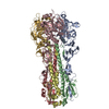
| |||||||||||||||||||||||||||||||||||||||||||||||||||||||||||||||||||||||||||||||||||||||||||||||||||||||||||||||||||||||||||||||||||||||||||||||||||||||||||||||||||||||||||||||||||||||||||||||||||||||||||||||||||||
| 2 | 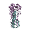
| |||||||||||||||||||||||||||||||||||||||||||||||||||||||||||||||||||||||||||||||||||||||||||||||||||||||||||||||||||||||||||||||||||||||||||||||||||||||||||||||||||||||||||||||||||||||||||||||||||||||||||||||||||||
| Unit cell |
| |||||||||||||||||||||||||||||||||||||||||||||||||||||||||||||||||||||||||||||||||||||||||||||||||||||||||||||||||||||||||||||||||||||||||||||||||||||||||||||||||||||||||||||||||||||||||||||||||||||||||||||||||||||
| Noncrystallographic symmetry (NCS) | NCS domain:
NCS domain segments: Component-ID: _ / Refine code: _
|