 Movie
Movie Controller

[English] 日本語
 Yorodumi
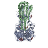
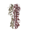
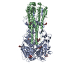
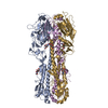
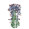
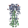
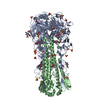
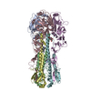
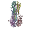
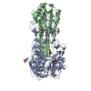
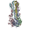
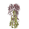
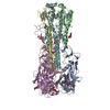
- PDB-6d7u: The crystal structure of hemagglutinin from A/Guangdong/17SF003/2... -
+ Open data

- Basic information
| Entry | Database: PDB / ID: 6d7u | ||||||
|---|---|---|---|---|---|---|---|
| Title | The crystal structure of hemagglutinin from A/Guangdong/17SF003/2016 H7N9 influenza virus | ||||||
 Components Components |
| ||||||
 Keywords Keywords | VIRAL PROTEIN / influenza / H7N9 | ||||||
| Function / homology |  Function and homology information Function and homology informationviral budding from plasma membrane / clathrin-dependent endocytosis of virus by host cell / apical plasma membrane / host cell surface receptor binding / fusion of virus membrane with host plasma membrane / fusion of virus membrane with host endosome membrane / viral envelope / virion attachment to host cell / host cell plasma membrane / virion membrane Similarity search - Function | ||||||
| Biological species |   Influenza A virus Influenza A virus | ||||||
| Method |  X-RAY DIFFRACTION / SYNCHROTRON / MOLECULAR REPLACEMENT / Resolution: 2.7 Å X-RAY DIFFRACTION / SYNCHROTRON / MOLECULAR REPLACEMENT / Resolution: 2.7 Å | ||||||
 Authors Authors | Yang, H. / Stevens, J. | ||||||
 Citation Citation | Journal: J. Virol. / Year: 2018 Title: Structural and Molecular Characterization of the Hemagglutinin from the Fifth-Epidemic-Wave A(H7N9) Influenza Viruses. Authors: Yang, H. / Carney, P.J. / Chang, J.C. / Guo, Z. / Stevens, J. | ||||||
| History |
|
- Structure visualization
| Structure viewer | Molecule: MolmilJmol/JSmol |
|---|
- Downloads & links
-Download
| PDBx/mmCIF format | 6d7u.cif.gz | 293.7 KB | Display | PDBx/mmCIF format |
|---|---|---|---|---|
| PDB format | pdb6d7u.ent.gz | 240.6 KB | Display | PDB format |
| PDBx/mmJSON format | 6d7u.json.gz | Tree view | PDBx/mmJSON format | |
| Others |  Other downloads Other downloads |
-Validation report
| Arichive directory | https://data.pdbj.org/pub/pdb/validation_reports/d7/6d7uftp://data.pdbj.org/pub/pdb/validation_reports/d7/6d7u | HTTPS FTP |
|---|
-Related structure data
| Related structure data |  6d7cC  6d8bC  6d8dC 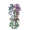 4ln6S S: Starting model for refinement C: citing same article ( |
|---|---|
| Similar structure data |


-Links
 PDBj
PDBj


- Assembly
| Deposited unit | 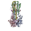
| ||||||||
|---|---|---|---|---|---|---|---|---|---|
| 1 |
| ||||||||
| Unit cell |
|
-Components
| #1: Protein | Mass: 34577.883 Da / Num. of mol.: 3 Source method: isolated from a genetically manipulated source Source: (gene. exp.) Influenza A virus / Strain: A/Guangdong/17SF003/2016(H7N9) / Gene: HA / Production host:  Trichoplusia ni (cabbage looper) / References: UniProt: A0A1S6R2B6 Trichoplusia ni (cabbage looper) / References: UniProt: A0A1S6R2B6#2: Protein | Mass: 19772.805 Da / Num. of mol.: 3 Source method: isolated from a genetically manipulated source Source: (gene. exp.) Influenza A virus / Strain: A/Guangdong/17SF003/2016(H7N9) / Gene: HA / Production host: Trichoplusia ni (cabbage looper) / References: UniProt: A0A2I7YV20#3: Sugar | ChemComp-NAG /   Type: D-saccharide, beta linking / Mass: 221.208 Da / Num. of mol.: 9 Type: D-saccharide, beta linking / Mass: 221.208 Da / Num. of mol.: 9Source method: isolated from a genetically manipulated source Formula: C8H15NO6 Has protein modification | Y | |
|---|
-Experimental details
-Experiment
| Experiment | Method: X-RAY DIFFRACTION / Number of used crystals: 1 |
|---|
- Sample preparation
| Crystal | Density Matthews: 3.61 Å3/Da / Density % sol: 65.89 % |
|---|---|
| Crystal grow | Temperature: 293 K / Method: microbatch / Details: 0.2M Ammonium Formate, pH 6.6, 20% PEG 3350 |
-Data collection
| Diffraction | Mean temperature: 100 K |
|---|---|
| Diffraction source | Source: SYNCHROTRON / Site: APS  / Beamline: 22-BM / Wavelength: 1 Å / Beamline: 22-BM / Wavelength: 1 Å |
| Detector | Type: MARMOSAIC 225 mm CCD / Detector: CCD / Date: Aug 7, 2017 |
| Radiation | Protocol: SINGLE WAVELENGTH / Monochromatic (M) / Laue (L): M / Scattering type: x-ray |
| Radiation wavelength | Wavelength: 1 Å / Relative weight: 1 |
| Reflection | Resolution: 2.7→50 Å / Num. obs: 62809 / % possible obs: 98.65 % / Redundancy: 3.2 % / Net I/σ(I): 22.27 |
| Reflection shell | Resolution: 2.7→2.75 Å |
- Processing
| Software |
| ||||||||||||||||||||||||||||||||||||||||||||||||||||||||||||||||||||||||||||||||||||||||||||||||||||||||||||||||||||||||||||||||||||||||||||||||||||||||||||||||||||||||
|---|---|---|---|---|---|---|---|---|---|---|---|---|---|---|---|---|---|---|---|---|---|---|---|---|---|---|---|---|---|---|---|---|---|---|---|---|---|---|---|---|---|---|---|---|---|---|---|---|---|---|---|---|---|---|---|---|---|---|---|---|---|---|---|---|---|---|---|---|---|---|---|---|---|---|---|---|---|---|---|---|---|---|---|---|---|---|---|---|---|---|---|---|---|---|---|---|---|---|---|---|---|---|---|---|---|---|---|---|---|---|---|---|---|---|---|---|---|---|---|---|---|---|---|---|---|---|---|---|---|---|---|---|---|---|---|---|---|---|---|---|---|---|---|---|---|---|---|---|---|---|---|---|---|---|---|---|---|---|---|---|---|---|---|---|---|---|---|---|---|
| Refinement | Method to determine structure: MOLECULAR REPLACEMENT Starting model: 4LN6 Resolution: 2.7→37.998 Å / SU ML: 0.39 / Cross valid method: FREE R-VALUE / σ(F): 1.37 / Phase error: 27.97
| ||||||||||||||||||||||||||||||||||||||||||||||||||||||||||||||||||||||||||||||||||||||||||||||||||||||||||||||||||||||||||||||||||||||||||||||||||||||||||||||||||||||||
| Solvent computation | Shrinkage radii: 0.9 Å / VDW probe radii: 1.11 Å | ||||||||||||||||||||||||||||||||||||||||||||||||||||||||||||||||||||||||||||||||||||||||||||||||||||||||||||||||||||||||||||||||||||||||||||||||||||||||||||||||||||||||
| Refinement step | Cycle: LAST / Resolution: 2.7→37.998 Å
| ||||||||||||||||||||||||||||||||||||||||||||||||||||||||||||||||||||||||||||||||||||||||||||||||||||||||||||||||||||||||||||||||||||||||||||||||||||||||||||||||||||||||
| Refine LS restraints |
| ||||||||||||||||||||||||||||||||||||||||||||||||||||||||||||||||||||||||||||||||||||||||||||||||||||||||||||||||||||||||||||||||||||||||||||||||||||||||||||||||||||||||
| LS refinement shell |
|
