 Movie
Movie Controller

[English] 日本語
 Yorodumi
- PDB-6n2e: Crystal Structure of Human Protocadherin-15 EC1-3 G16D N369D Q370... -
+ Open data

- Basic information
| Entry | Database: PDB / ID: 6n2e | ||||||
|---|---|---|---|---|---|---|---|
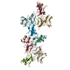
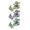
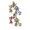
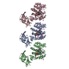
| Title | Crystal Structure of Human Protocadherin-15 EC1-3 G16D N369D Q370N and Mouse Cadherin-23 EC1-2 T15E | ||||||
 Components Components |
| ||||||
 Keywords Keywords | CELL ADHESION / Mechanotransduction / Calcium-binding protein / stereocilia / hair cell / tip link | ||||||
| Function / homology |  Function and homology information Function and homology informationcochlear hair cell ribbon synapse / equilibrioception / sensory perception of light stimulus / stereocilium tip / inner ear receptor cell stereocilium organization / photoreceptor ribbon synapse / kinocilium / inner ear auditory receptor cell differentiation / : / calcium-dependent cell-cell adhesion ...cochlear hair cell ribbon synapse / equilibrioception / sensory perception of light stimulus / stereocilium tip / inner ear receptor cell stereocilium organization / photoreceptor ribbon synapse / kinocilium / inner ear auditory receptor cell differentiation / : / calcium-dependent cell-cell adhesion / catenin complex / stereocilium / photoreceptor cell maintenance / auditory receptor cell stereocilium organization / Sensory processing of sound by outer hair cells of the cochlea / Sensory processing of sound by inner hair cells of the cochlea / inner ear morphogenesis / homophilic cell-cell adhesion / inner ear development / cochlea development / photoreceptor outer segment / regulation of cytosolic calcium ion concentration / photoreceptor inner segment / cell adhesion molecule binding / locomotory behavior / sensory perception of sound / beta-catenin binding / apical part of cell / neuron projection development / calcium ion transport / cell migration / cell adhesion / cadherin binding / calcium ion binding / centrosome / synapse / : / extracellular region / plasma membrane Similarity search - Function | ||||||
| Biological species |  Homo sapiens (human) Homo sapiens (human) | ||||||
| Method |  X-RAY DIFFRACTION / SYNCHROTRON / MOLECULAR REPLACEMENT / Resolution: 2.9 Å X-RAY DIFFRACTION / SYNCHROTRON / MOLECULAR REPLACEMENT / Resolution: 2.9 Å | ||||||
 Authors Authors | Choudhary, D. / De-la-Torre, P. / Sotomayor, M. | ||||||
| Funding support |  United States, 1items United States, 1items
| ||||||
 Citation Citation | Journal: Proc.Natl.Acad.Sci.USA / Year: 2020 Title: Structural determinants of protocadherin-15 mechanics and function in hearing and balance perception. Authors: Choudhary, D. / Narui, Y. / Neel, B.L. / Wimalasena, L.N. / Klanseck, C.F. / De-la-Torre, P. / Chen, C. / Araya-Secchi, R. / Tamilselvan, E. / Sotomayor, M. | ||||||
| History |
|
- Structure visualization
| Structure viewer | Molecule: MolmilJmol/JSmol |
|---|
- Downloads & links
-Download
| PDBx/mmCIF format | 6n2e.cif.gz | 452.7 KB | Display | PDBx/mmCIF format |
|---|---|---|---|---|
| PDB format | pdb6n2e.ent.gz | 372.7 KB | Display | PDB format |
| PDBx/mmJSON format | 6n2e.json.gz | Tree view | PDBx/mmJSON format | |
| Others |  Other downloads Other downloads |
-Validation report
| Arichive directory | https://data.pdbj.org/pub/pdb/validation_reports/n2/6n2eftp://data.pdbj.org/pub/pdb/validation_reports/n2/6n2e | HTTPS FTP |
|---|
-Related structure data
| Related structure data | 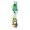 5tpkC 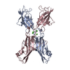 5ulyC 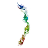 5w1dC 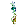 6bwnC  6bxuC  6e8fC  6eb5C  6eetC  6mfoSC  6n22C  4aq8S S: Starting model for refinement C: citing same article ( |
|---|---|
| Similar structure data |

-Links
 PDBj
PDBj


- Assembly
| Deposited unit | 
| ||||||||
|---|---|---|---|---|---|---|---|---|---|
| 1 |
| ||||||||
| Unit cell |
|
-Components
| #1: Protein | Mass: 43069.160 Da / Num. of mol.: 2 / Mutation: G16D, N369D, Q370N Source method: isolated from a genetically manipulated source Source: (gene. exp.) Homo sapiens (human) / Gene: PCDH15 / Plasmid: pET21a / Production host:  #2: Protein | Mass: 23884.445 Da / Num. of mol.: 2 / Mutation: T15E Source method: isolated from a genetically manipulated source Source: (gene. exp.) #3: Chemical | ChemComp-CA /   Mass: 40.078 Da / Num. of mol.: 18 / Source method: obtained synthetically / Formula: Ca Mass: 40.078 Da / Num. of mol.: 18 / Source method: obtained synthetically / Formula: Ca#4: Water | ChemComp-HOH / |  Mass: 18.015 Da / Num. of mol.: 62 / Source method: isolated from a natural source / Formula: H2O Mass: 18.015 Da / Num. of mol.: 62 / Source method: isolated from a natural source / Formula: H2OHas protein modification | Y | |
|---|
-Experimental details
-Experiment
| Experiment | Method: X-RAY DIFFRACTION / Number of used crystals: 1 |
|---|
- Sample preparation
| Crystal | Density Matthews: 3.52 Å3/Da / Density % sol: 65.08 % |
|---|---|
| Crystal grow | Temperature: 277 K / Method: vapor diffusion, sitting drop / Details: 0.1 M Imidazole pH 6.8, 46% MPD |
-Data collection
| Diffraction | Mean temperature: 100 K / Serial crystal experiment: N |
|---|---|
| Diffraction source | Source: SYNCHROTRON / Site: APS / Beamline: 24-ID-E / Wavelength: 0.97918 Å |
| Detector | Type: DECTRIS EIGER X 16M / Detector: PIXEL / Date: Oct 18, 2018 |
| Radiation | Protocol: SINGLE WAVELENGTH / Monochromatic (M) / Laue (L): M / Scattering type: x-ray |
| Radiation wavelength | Wavelength: 0.97918 Å / Relative weight: 1 |
| Reflection | Resolution: 2.9→50 Å / Num. obs: 41847 / % possible obs: 93.2 % / Redundancy: 5.2 % / Rmerge(I) obs: 0.115 / Rpim(I) all: 0.055 / Net I/σ(I): 12.33 |
| Reflection shell | Resolution: 2.9→2.95 Å / Redundancy: 4.4 % / Num. unique obs: 1806 / Rpim(I) all: 0.236 / % possible all: 86.5 |
- Processing
| Software |
| ||||||||||||||||||||||||||||||||||||||||||||||||||||||||||||||||||||||||||||||||||||||||||||||||||||||||||||||||||||||||||||||||||||||||||||||||||||||||||||||||||||||||||||||||||||||
|---|---|---|---|---|---|---|---|---|---|---|---|---|---|---|---|---|---|---|---|---|---|---|---|---|---|---|---|---|---|---|---|---|---|---|---|---|---|---|---|---|---|---|---|---|---|---|---|---|---|---|---|---|---|---|---|---|---|---|---|---|---|---|---|---|---|---|---|---|---|---|---|---|---|---|---|---|---|---|---|---|---|---|---|---|---|---|---|---|---|---|---|---|---|---|---|---|---|---|---|---|---|---|---|---|---|---|---|---|---|---|---|---|---|---|---|---|---|---|---|---|---|---|---|---|---|---|---|---|---|---|---|---|---|---|---|---|---|---|---|---|---|---|---|---|---|---|---|---|---|---|---|---|---|---|---|---|---|---|---|---|---|---|---|---|---|---|---|---|---|---|---|---|---|---|---|---|---|---|---|---|---|---|---|
| Refinement | Method to determine structure: MOLECULAR REPLACEMENT Starting model: 6MFO, 4AQ8 Resolution: 2.9→49.52 Å / Cor.coef. Fo:Fc: 0.923 / Cor.coef. Fo:Fc free: 0.908 / SU B: 22.739 / SU ML: 0.211 / Cross valid method: THROUGHOUT / ESU R: 0.305 / ESU R Free: 0.079 / Details: HYDROGENS HAVE BEEN ADDED IN THE RIDING POSITIONS
| ||||||||||||||||||||||||||||||||||||||||||||||||||||||||||||||||||||||||||||||||||||||||||||||||||||||||||||||||||||||||||||||||||||||||||||||||||||||||||||||||||||||||||||||||||||||
| Solvent computation | Ion probe radii: 0.9 Å / Shrinkage radii: 0.9 Å / VDW probe radii: 1.1 Å | ||||||||||||||||||||||||||||||||||||||||||||||||||||||||||||||||||||||||||||||||||||||||||||||||||||||||||||||||||||||||||||||||||||||||||||||||||||||||||||||||||||||||||||||||||||||
| Displacement parameters | Biso mean: 75.813 Å2
| ||||||||||||||||||||||||||||||||||||||||||||||||||||||||||||||||||||||||||||||||||||||||||||||||||||||||||||||||||||||||||||||||||||||||||||||||||||||||||||||||||||||||||||||||||||||
| Refinement step | Cycle: 1 / Resolution: 2.9→49.52 Å
| ||||||||||||||||||||||||||||||||||||||||||||||||||||||||||||||||||||||||||||||||||||||||||||||||||||||||||||||||||||||||||||||||||||||||||||||||||||||||||||||||||||||||||||||||||||||
| Refine LS restraints |
|