Movie
Movie Controller

[English] 日本語
 Yorodumi
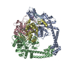
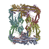
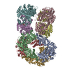
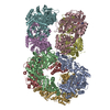
- PDB-6n1p: Dihedral oligomeric complex of GyrA N-terminal fragment with DNA,... -
+ Open data

- Basic information
| Entry | Database: PDB / ID: 6n1p | ||||||||||||||||||
|---|---|---|---|---|---|---|---|---|---|---|---|---|---|---|---|---|---|---|---|
| Title | Dihedral oligomeric complex of GyrA N-terminal fragment with DNA, solved by cryoEM in C2 symmetry | ||||||||||||||||||
 Components Components |
| ||||||||||||||||||
 Keywords Keywords | ISOMERASE/DNA / topoisomerase / oligomeric complex / DNA complex / gyrase / T-segment / ISOMERASE-DNA complex | ||||||||||||||||||
| Function / homology |  Function and homology information Function and homology informationDNA topoisomerase type II (double strand cut, ATP-hydrolyzing) complex / DNA negative supercoiling activity / DNA topoisomerase type II (double strand cut, ATP-hydrolyzing) activity / DNA topoisomerase (ATP-hydrolysing) / DNA topological change / DNA-templated DNA replication / chromosome / DNA binding / ATP binding / cytoplasm Similarity search - Function | ||||||||||||||||||
| Biological species |  Streptococcus pneumoniae G54 (bacteria) Streptococcus pneumoniae G54 (bacteria)Cloning vector pBR322 (others) | ||||||||||||||||||
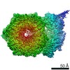
| Method | ELECTRON MICROSCOPY / single particle reconstruction / cryo EM / Resolution: 6.35 Å | ||||||||||||||||||
 Authors Authors | Soczek, K.M. / Grant, T. / Rosenthal, P.B. / Mondragon, A. | ||||||||||||||||||
| Funding support |  United States, United States,  United Kingdom, 5items United Kingdom, 5items
| ||||||||||||||||||
 Citation Citation | Journal: Elife / Year: 2018 Title: CryoEM structures of open dimers of gyrase A in complex with DNA illuminate mechanism of strand passage. Authors: Katarzyna M Soczek / Tim Grant / Peter B Rosenthal / Alfonso Mondragón / Abstract: Gyrase is a unique type IIA topoisomerase that uses ATP hydrolysis to maintain the negatively supercoiled state of bacterial DNA. In order to perform its function, gyrase undergoes a sequence of ...Gyrase is a unique type IIA topoisomerase that uses ATP hydrolysis to maintain the negatively supercoiled state of bacterial DNA. In order to perform its function, gyrase undergoes a sequence of conformational changes that consist of concerted gate openings, DNA cleavage, and DNA strand passage events. Structures where the transported DNA molecule (T-segment) is trapped by the A subunit have not been observed. Here we present the cryoEM structures of two oligomeric complexes of open gyrase A dimers and DNA. The protein subunits in these complexes were solved to 4 Å and 5.2 Å resolution. One of the complexes traps a linear DNA molecule, a putative T-segment, which interacts with the open gyrase A dimers in two states, representing steps either prior to or after passage through the DNA-gate. The structures locate the T-segment in important intermediate conformations of the catalytic cycle and provide insights into gyrase-DNA interactions and mechanism. | ||||||||||||||||||
| History |
|
- Structure visualization
| Movie |
Movie viewer |
|---|---|
| Structure viewer | Molecule: MolmilJmol/JSmol |
- Downloads & links
-Download
| PDBx/mmCIF format | 6n1p.cif.gz | 801.8 KB | Display | PDBx/mmCIF format |
|---|---|---|---|---|
| PDB format | pdb6n1p.ent.gz | 658.5 KB | Display | PDB format |
| PDBx/mmJSON format | 6n1p.json.gz | Tree view | PDBx/mmJSON format | |
| Others |  Other downloads Other downloads |
-Validation report
| Arichive directory | https://data.pdbj.org/pub/pdb/validation_reports/n1/6n1pftp://data.pdbj.org/pub/pdb/validation_reports/n1/6n1p | HTTPS FTP |
|---|
-Related structure data
| Related structure data |  9316MC 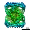 9317C 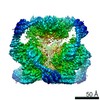 9318C 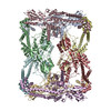 6n1qC 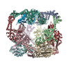 6n1rC M: map data used to model this data C: citing same article ( |
|---|---|
| Similar structure data |
-Links
 PDBj
PDBj


- Assembly
| Deposited unit | 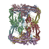
|
|---|---|
| 1 |
|
-Components
| #1: Protein | Mass: 57977.578 Da / Num. of mol.: 8 / Fragment: UNP residues 20-506 Source method: isolated from a genetically manipulated source Source: (gene. exp.) Streptococcus pneumoniae G54 (bacteria)Gene: gyrA_1, gyrA, ERS409327_01712 / Plasmid: pMCSG7 / Production host: References: UniProt: A0A0Y2BJX7, UniProt: Q8DPM2*PLUS, EC: 5.99.1.3 #2: DNA chain | | Mass: 13641.771 Da / Num. of mol.: 1 / Source method: obtained synthetically / Source: (synth.) Cloning vector pBR322 (others) #3: DNA chain | | Mass: 13458.600 Da / Num. of mol.: 1 / Source method: obtained synthetically / Source: (synth.) Cloning vector pBR322 (others) |
|---|
-Experimental details
-Experiment
| Experiment | Method: ELECTRON MICROSCOPY |
|---|---|
| EM experiment | Aggregation state: PARTICLE / 3D reconstruction method: single particle reconstruction |
- Sample preparation
| Component |
| ||||||||||||||||||||||||
|---|---|---|---|---|---|---|---|---|---|---|---|---|---|---|---|---|---|---|---|---|---|---|---|---|---|
| Molecular weight | Value: 0.49 MDa / Experimental value: NO | ||||||||||||||||||||||||
| Source (natural) |
| ||||||||||||||||||||||||
| Source (recombinant) | Organism: | ||||||||||||||||||||||||
| Buffer solution | pH: 7.5 | ||||||||||||||||||||||||
| Buffer component |
| ||||||||||||||||||||||||
| Specimen | Embedding applied: NO / Shadowing applied: NO / Staining applied: NO / Vitrification applied: YES | ||||||||||||||||||||||||
| Specimen support | Grid material: COPPER / Grid mesh size: 400 divisions/in. / Grid type: C-flat-1.2/1.3 4C | ||||||||||||||||||||||||
| Vitrification | Instrument: FEI VITROBOT MARK IV / Cryogen name: ETHANE / Humidity: 100 % / Chamber temperature: 277 K |
- Electron microscopy imaging
| Microscopy | Model: JEOL 3200FS |
|---|---|
| Electron gun | Electron source:  FIELD EMISSION GUN / Accelerating voltage: 300 kV / Illumination mode: OTHER FIELD EMISSION GUN / Accelerating voltage: 300 kV / Illumination mode: OTHER |
| Electron lens | Mode: BRIGHT FIELD / Nominal magnification: 30000 X / Calibrated magnification: 40323 X / Nominal defocus max: 3000 nm / Nominal defocus min: 1000 nm / Cs: 2 mm / C2 aperture diameter: 50 µm / Alignment procedure: COMA FREE |
| Specimen holder | Cryogen: NITROGEN Specimen holder model: GATAN 626 SINGLE TILT LIQUID NITROGEN CRYO TRANSFER HOLDER Temperature (max): 100 K / Temperature (min): 100 K |
| Image recording | Average exposure time: 12 sec. / Electron dose: 62 e/Å2 / Detector mode: COUNTING / Film or detector model: GATAN K2 SUMMIT (4k x 4k) / Num. of real images: 718 |
| EM imaging optics | Energyfilter name: In-column Omega Filter / Energyfilter slit width: 20 eV |
| Image scans | Sampling size: 5 µm / Width: 3710 / Height: 3838 / Movie frames/image: 40 / Used frames/image: 1-40 |
- Processing
| EM software |
| ||||||||||||||||||||||||||||||||||||||||||||
|---|---|---|---|---|---|---|---|---|---|---|---|---|---|---|---|---|---|---|---|---|---|---|---|---|---|---|---|---|---|---|---|---|---|---|---|---|---|---|---|---|---|---|---|---|---|
| CTF correction | Type: PHASE FLIPPING AND AMPLITUDE CORRECTION | ||||||||||||||||||||||||||||||||||||||||||||
| Particle selection | Num. of particles selected: 387297 | ||||||||||||||||||||||||||||||||||||||||||||
| Symmetry | Point symmetry: C2 (2 fold cyclic) | ||||||||||||||||||||||||||||||||||||||||||||
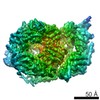
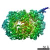
| 3D reconstruction | Resolution: 6.35 Å / Resolution method: FSC 0.143 CUT-OFF / Num. of particles: 30637 / Algorithm: FOURIER SPACE / Num. of class averages: 1 / Symmetry type: POINT | ||||||||||||||||||||||||||||||||||||||||||||
| Atomic model building | Protocol: FLEXIBLE FIT / Space: REAL / Target criteria: correlation coefficient | ||||||||||||||||||||||||||||||||||||||||||||
| Atomic model building | PDB-ID: 4Z2C Pdb chain-ID: A / Accession code: 4Z2C / Pdb chain residue range: 2-486 / Source name: PDB / Type: experimental model |