Movie
Movie Controller

[English] 日本語
 Yorodumi
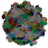
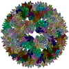
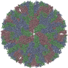
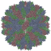
- PDB-6mzy: Cryo-EM structure of the HO BMC shell: Icosahedral reconstruction... -
+ Open data

- Basic information
| Entry | Database: PDB / ID: 6mzy | |||||||||
|---|---|---|---|---|---|---|---|---|---|---|
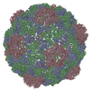
| Title | Cryo-EM structure of the HO BMC shell: Icosahedral reconstruction of the compacted subpopulation | |||||||||
 Components Components |
| |||||||||
 Keywords Keywords | STRUCTURAL PROTEIN / microcompartment / shell / compartmentalization / BMC fold | |||||||||
| Function / homology |  Function and homology information Function and homology information | |||||||||
| Biological species |  Haliangium ochraceum (bacteria) Haliangium ochraceum (bacteria) | |||||||||
| Method | ELECTRON MICROSCOPY / single particle reconstruction / cryo EM / Resolution: 3.3 Å | |||||||||
 Authors Authors | Greber, B.J. / Sutter, M. / Kerfeld, C.A. | |||||||||
| Funding support |  United States, 2items United States, 2items
| |||||||||
 Citation Citation | Journal: Structure / Year: 2019 Title: The Plasticity of Molecular Interactions Governs Bacterial Microcompartment Shell Assembly. Authors: Basil J Greber / Markus Sutter / Cheryl A Kerfeld / Abstract: Bacterial microcompartments (BMCs) are composed of an enzymatic core encapsulated by a selectively permeable protein shell that enhances catalytic efficiency. Many pathogenic bacteria derive ...Bacterial microcompartments (BMCs) are composed of an enzymatic core encapsulated by a selectively permeable protein shell that enhances catalytic efficiency. Many pathogenic bacteria derive competitive advantages from their BMC-based catabolism, implicating BMCs as drug targets. BMC shells are of interest for bioengineering due to their diverse and selective permeability properties and because they self-assemble. A complete understanding of shell composition and organization is a prerequisite for biotechnological applications. Here, we report the cryoelectron microscopy structure of a BMC shell at 3.0-Å resolution, using an image-processing strategy that allowed us to determine the previously uncharacterized structural details of the interactions formed by the BMC-T and BMC-T shell subunits in the context of the assembled shell. We found unexpected structural plasticity among these interactions, resulting in distinct shell populations assembled from varying numbers of the BMC-T and BMC-T subunits. We discuss the implications of these findings on shell assembly and function. | |||||||||
| History |
|
- Structure visualization
| Movie |
Movie viewer |
|---|---|
| Structure viewer | Molecule: MolmilJmol/JSmol |
- Downloads & links
-Download
| PDBx/mmCIF format | 6mzy.cif.gz | 170.3 KB | Display | PDBx/mmCIF format |
|---|---|---|---|---|
| PDB format | pdb6mzy.ent.gz | Display | PDB format | |
| PDBx/mmJSON format | 6mzy.json.gz | Tree view | PDBx/mmJSON format | |
| Others |  Other downloads Other downloads |
-Validation report
| Arichive directory | https://data.pdbj.org/pub/pdb/validation_reports/mz/6mzyftp://data.pdbj.org/pub/pdb/validation_reports/mz/6mzy | HTTPS FTP |
|---|
-Related structure data
| Related structure data | 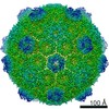 9310MC 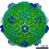 9296C 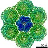 9307C 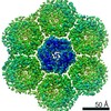 9308C 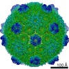 9309C 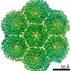 9311C 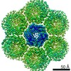 9312C 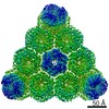 9313C 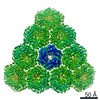 9314C 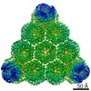 9315C 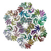 6mzuC 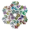 6mzvC 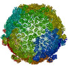 6mzxC 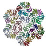 6n06C 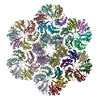 6n07C 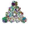 6n09C  6n0fC  6n0gC M: map data used to model this data C: citing same article ( |
|---|---|
| Similar structure data |

-Links
 PDBj
PDBj

- Assembly
| Deposited unit | 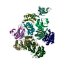
|
|---|---|
| 1 | x 60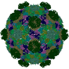
|
| 2 |
|
| 3 | x 5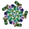
|
| 4 | x 6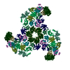
|
| 5 | 
|
| Symmetry | Point symmetry: (Schoenflies symbol: I (icosahedral)) |
-Components
| #1: Protein | Mass: 9866.319 Da / Num. of mol.: 1 Source method: isolated from a genetically manipulated source Source: (gene. exp.) Haliangium ochraceum (strain DSM 14365 / JCM 11303 / SMP-2) (bacteria)Strain: DSM 14365 / JCM 11303 / SMP-2 / Gene: Hoch_5814 / Production host: | ||
|---|---|---|---|
| #2: Protein | Mass: 10126.718 Da / Num. of mol.: 6 Source method: isolated from a genetically manipulated source Source: (gene. exp.) Haliangium ochraceum (strain DSM 14365 / JCM 11303 / SMP-2) (bacteria)Strain: DSM 14365 / JCM 11303 / SMP-2 / Gene: Hoch_5815 / Production host: #3: Protein | Mass: 22904.137 Da / Num. of mol.: 2 Source method: isolated from a genetically manipulated source Source: (gene. exp.) Haliangium ochraceum (strain DSM 14365 / JCM 11303 / SMP-2) (bacteria)Strain: DSM 14365 / JCM 11303 / SMP-2 / Gene: Hoch_5816 / Production host: |
-Experimental details
-Experiment
| Experiment | Method: ELECTRON MICROSCOPY |
|---|---|
| EM experiment | Aggregation state: PARTICLE / 3D reconstruction method: single particle reconstruction |
- Sample preparation
| Component | Name: Bacterial microcompartment shell from Haliangium ochraceum Type: ORGANELLE OR CELLULAR COMPONENT / Entity ID: all / Source: RECOMBINANT | ||||||||||||||||||||
|---|---|---|---|---|---|---|---|---|---|---|---|---|---|---|---|---|---|---|---|---|---|
| Molecular weight | Value: 6.5 MDa / Experimental value: NO | ||||||||||||||||||||
| Source (natural) | Organism: Haliangium ochraceum (bacteria) | ||||||||||||||||||||
| Source (recombinant) | Organism: | ||||||||||||||||||||
| Buffer solution | pH: 7.4 | ||||||||||||||||||||
| Buffer component |
| ||||||||||||||||||||
| Specimen | Conc.: 3 mg/ml / Embedding applied: NO / Shadowing applied: NO / Staining applied: NO / Vitrification applied: YES | ||||||||||||||||||||
| Specimen support | Details: unspecified | ||||||||||||||||||||
| Vitrification | Instrument: FEI VITROBOT MARK IV / Cryogen name: ETHANE / Humidity: 100 % / Chamber temperature: 277 K Details: 5-7 sec incubation of the sample on the grid before blotting and plunging |
- Electron microscopy imaging
| Microscopy | Model: FEI TITAN |
|---|---|
| Electron gun | Electron source:  FIELD EMISSION GUN / Accelerating voltage: 300 kV / Illumination mode: FLOOD BEAM FIELD EMISSION GUN / Accelerating voltage: 300 kV / Illumination mode: FLOOD BEAM |
| Electron lens | Mode: BRIGHT FIELD / Calibrated magnification: 48543 X / Calibrated defocus min: 1000 nm / Calibrated defocus max: 3500 nm / Cs: 2.7 mm / C2 aperture diameter: 50 µm / Alignment procedure: COMA FREE |
| Specimen holder | Cryogen: NITROGEN Specimen holder model: GATAN 626 SINGLE TILT LIQUID NITROGEN CRYO TRANSFER HOLDER |
| Image recording | Average exposure time: 4.5 sec. / Electron dose: 25 e/Å2 / Detector mode: COUNTING / Film or detector model: GATAN K2 SUMMIT (4k x 4k) / Num. of grids imaged: 1 / Num. of real images: 928 Details: 928 images retained after inspection for image quality. Movie frames were aligned and dose weighed using Motioncor2. |
| Image scans | Sampling size: 5 µm / Width: 3838 / Height: 3710 / Movie frames/image: 30 / Used frames/image: 1-30 |
- Processing
| EM software |
| ||||||||||||||||||||||||||||||||||||||||
|---|---|---|---|---|---|---|---|---|---|---|---|---|---|---|---|---|---|---|---|---|---|---|---|---|---|---|---|---|---|---|---|---|---|---|---|---|---|---|---|---|---|
| CTF correction | Details: Initial CTF fitting using CTFFIND4, CTF correction applied within RELION. Type: PHASE FLIPPING AND AMPLITUDE CORRECTION | ||||||||||||||||||||||||||||||||||||||||
| Particle selection | Num. of particles selected: 31800 Details: 1000 particles were picked manually to generate reference templates for subsequent auto-picking in RELION 1.4. | ||||||||||||||||||||||||||||||||||||||||
| Symmetry | Point symmetry: I (icosahedral) | ||||||||||||||||||||||||||||||||||||||||
| 3D reconstruction | Resolution: 3.3 Å / Resolution method: FSC 0.143 CUT-OFF / Num. of particles: 2276 / Algorithm: FOURIER SPACE Details: Frequency-limited refinement in FREALIGN. To avoid overfitting, resolutions higher than 3.6 Angstroms were excluded from the refinement. Icosahedral symmetry was imposed throughout the refinement procedure. Num. of class averages: 1 / Symmetry type: POINT | ||||||||||||||||||||||||||||||||||||||||
| Atomic model building | Protocol: OTHER / Space: REAL | ||||||||||||||||||||||||||||||||||||||||
| Atomic model building | PDB-ID: 5V74 Accession code: 5V74 / Source name: PDB / Type: experimental model |