 Movie
Movie Controller

[English] 日本語
 Yorodumi
- PDB-6myn: Crystal structure of murine NF-kappaB inducing kinase (NIK) bound... -
+ Open data

- Basic information
| Entry | Database: PDB / ID: 6myn | ||||||
|---|---|---|---|---|---|---|---|
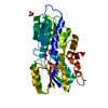
| Title | Crystal structure of murine NF-kappaB inducing kinase (NIK) bound to inhibitor R7 | ||||||
 Components Components | Mitogen-activated protein kinase kinase kinase 14 | ||||||
 Keywords Keywords | TRANSFERASE/TRANSFERASE Inhibitor / NIK / kinase / TRANSFERASE / TRANSFERASE-TRANSFERASE Inhibitor complex | ||||||
| Function / homology |  Function and homology information Function and homology informationTNF receptor superfamily (TNFSF) members mediating non-canonical NF-kB pathway / CD28 dependent PI3K/Akt signaling / TNFR2 non-canonical NF-kB pathway / Dectin-1 mediated noncanonical NF-kB signaling / NIK-->noncanonical NF-kB signaling / mitogen-activated protein kinase kinase kinase / non-canonical NF-kappaB signal transduction / MAP kinase kinase kinase activity / cellular response to mechanical stimulus / fibrillar center ...TNF receptor superfamily (TNFSF) members mediating non-canonical NF-kB pathway / CD28 dependent PI3K/Akt signaling / TNFR2 non-canonical NF-kB pathway / Dectin-1 mediated noncanonical NF-kB signaling / NIK-->noncanonical NF-kB signaling / mitogen-activated protein kinase kinase kinase / non-canonical NF-kappaB signal transduction / MAP kinase kinase kinase activity / cellular response to mechanical stimulus / fibrillar center / MAPK cascade / defense response to virus / protein kinase activity / immune response / protein serine kinase activity / protein serine/threonine kinase activity / nucleoplasm / ATP binding / cytosol Similarity search - Function | ||||||
| Biological species |  | ||||||
| Method |  X-RAY DIFFRACTION / SYNCHROTRON / FOURIER SYNTHESIS / Resolution: 2.744 Å X-RAY DIFFRACTION / SYNCHROTRON / FOURIER SYNTHESIS / Resolution: 2.744 Å | ||||||
 Authors Authors | Harris, S.F. / Smith, M. / Barker, J. | ||||||
 Citation Citation | Journal: Acs Med.Chem.Lett. / Year: 2019 Title: Structure Based Design of Potent Selective Inhibitors of Protein Kinase D1 (PKD1). Authors: Feng, J.A. / Lee, P. / Alaoui, M.H. / Barrett, K. / Castanedo, G. / Godemann, R. / McEwan, P. / Wang, X. / Wu, P. / Zhang, Y. / Harris, S.F. / Staben, S.T. #1: Journal: Acs Med.Chem.Lett. / Year: 2019Title: Structure based design of potent selective inhibitors of protein kinase D1 (PKD1) Authors: Feng, J.W. / Lee, P. / Alaoui, M.H. / Barrett, K. / Castaneda, G.M. / Godemann, R. / McEwan, P. / Wang, X. / Wu, P. / Zhang, Y. / Harris, S.F. / Staben, S.T. | ||||||
| History |
|
- Structure visualization
| Structure viewer | Molecule: MolmilJmol/JSmol |
|---|
- Downloads & links
-Download
| PDBx/mmCIF format | 6myn.cif.gz | 144.9 KB | Display | PDBx/mmCIF format |
|---|---|---|---|---|
| PDB format | pdb6myn.ent.gz | 112.9 KB | Display | PDB format |
| PDBx/mmJSON format | 6myn.json.gz | Tree view | PDBx/mmJSON format | |
| Others |  Other downloads Other downloads |
-Validation report
| Arichive directory | https://data.pdbj.org/pub/pdb/validation_reports/my/6mynftp://data.pdbj.org/pub/pdb/validation_reports/my/6myn | HTTPS FTP |
|---|
-Related structure data


-Links
 PDBj
PDBj
- Assembly
| Deposited unit | 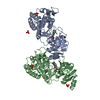
| ||||||||
|---|---|---|---|---|---|---|---|---|---|
| 1 | 
| ||||||||
| 2 | 
| ||||||||
| Unit cell |
|
-Components
| #1: Protein | Mass: 38399.320 Da / Num. of mol.: 2 Source method: isolated from a genetically manipulated source Source: (gene. exp.)   Spodoptera frugiperda (fall armyworm) Spodoptera frugiperda (fall armyworm)References: UniProt: Q9WUL6, mitogen-activated protein kinase kinase kinase #2: Chemical | ChemComp-SO4 /   Mass: 96.063 Da / Num. of mol.: 5 / Source method: obtained synthetically / Formula: SO4 Mass: 96.063 Da / Num. of mol.: 5 / Source method: obtained synthetically / Formula: SO4#3: Chemical |   Mass: 463.461 Da / Num. of mol.: 2 / Source method: obtained synthetically / Formula: C24H22FN5O4 / Feature type: SUBJECT OF INVESTIGATION Mass: 463.461 Da / Num. of mol.: 2 / Source method: obtained synthetically / Formula: C24H22FN5O4 / Feature type: SUBJECT OF INVESTIGATION#4: Water | ChemComp-HOH / |  Mass: 18.015 Da / Num. of mol.: 105 / Source method: isolated from a natural source / Formula: H2O Mass: 18.015 Da / Num. of mol.: 105 / Source method: isolated from a natural source / Formula: H2O |
|---|
-Experimental details
-Experiment
| Experiment | Method: X-RAY DIFFRACTION / Number of used crystals: 1 |
|---|
- Sample preparation
| Crystal | Density Matthews: 3.3 Å3/Da / Density % sol: 62.68 % |
|---|---|
| Crystal grow | Temperature: 277 K / Method: vapor diffusion, hanging drop / pH: 6.2 Details: 100 mM sodium citrate tribasic dihydrate (pH 6.2), 0.5 M ammonium sulfate, 0.9 M lithium sulfate |
-Data collection
| Diffraction | Mean temperature: 100 K / Serial crystal experiment: N | |||||||||||||||||||||||||||||||||||||||||||||||||||||||||||||||||||||||||||||||||||||||||||||||||||||||||||||||||||||||||||||||||||||||||||||||||||||||||||||||||||||||||||||||||||||||||||||
|---|---|---|---|---|---|---|---|---|---|---|---|---|---|---|---|---|---|---|---|---|---|---|---|---|---|---|---|---|---|---|---|---|---|---|---|---|---|---|---|---|---|---|---|---|---|---|---|---|---|---|---|---|---|---|---|---|---|---|---|---|---|---|---|---|---|---|---|---|---|---|---|---|---|---|---|---|---|---|---|---|---|---|---|---|---|---|---|---|---|---|---|---|---|---|---|---|---|---|---|---|---|---|---|---|---|---|---|---|---|---|---|---|---|---|---|---|---|---|---|---|---|---|---|---|---|---|---|---|---|---|---|---|---|---|---|---|---|---|---|---|---|---|---|---|---|---|---|---|---|---|---|---|---|---|---|---|---|---|---|---|---|---|---|---|---|---|---|---|---|---|---|---|---|---|---|---|---|---|---|---|---|---|---|---|---|---|---|---|---|---|
| Diffraction source | Source: SYNCHROTRON / Site: Diamond  / Beamline: I04-1 / Wavelength: 0.9173 Å / Beamline: I04-1 / Wavelength: 0.9173 Å | |||||||||||||||||||||||||||||||||||||||||||||||||||||||||||||||||||||||||||||||||||||||||||||||||||||||||||||||||||||||||||||||||||||||||||||||||||||||||||||||||||||||||||||||||||||||||||||
| Detector | Type: DECTRIS PILATUS 2M / Detector: PIXEL / Date: Aug 14, 2012 | |||||||||||||||||||||||||||||||||||||||||||||||||||||||||||||||||||||||||||||||||||||||||||||||||||||||||||||||||||||||||||||||||||||||||||||||||||||||||||||||||||||||||||||||||||||||||||||
| Radiation | Protocol: SINGLE WAVELENGTH / Monochromatic (M) / Laue (L): M / Scattering type: x-ray | |||||||||||||||||||||||||||||||||||||||||||||||||||||||||||||||||||||||||||||||||||||||||||||||||||||||||||||||||||||||||||||||||||||||||||||||||||||||||||||||||||||||||||||||||||||||||||||
| Radiation wavelength | Wavelength: 0.9173 Å / Relative weight: 1 | |||||||||||||||||||||||||||||||||||||||||||||||||||||||||||||||||||||||||||||||||||||||||||||||||||||||||||||||||||||||||||||||||||||||||||||||||||||||||||||||||||||||||||||||||||||||||||||
| Reflection | Resolution: 2.744→44.15 Å / Num. obs: 25142 / % possible obs: 99.9 % / Redundancy: 6.6 % / Biso Wilson estimate: 58.27 Å2 / Rpim(I) all: 0.041 / Rrim(I) all: 0.106 / Rsym value: 0.086 / Net I/av σ(I): 8.4 / Net I/σ(I): 16 | |||||||||||||||||||||||||||||||||||||||||||||||||||||||||||||||||||||||||||||||||||||||||||||||||||||||||||||||||||||||||||||||||||||||||||||||||||||||||||||||||||||||||||||||||||||||||||||
| Reflection shell | Diffraction-ID: 1
|
- Processing
| Software |
| ||||||||||||||||||||||||||||||||||||||||||||||||||||||||||||||||||||||||||||||||||||||||||||||||||||||||||||||||||||||||
|---|---|---|---|---|---|---|---|---|---|---|---|---|---|---|---|---|---|---|---|---|---|---|---|---|---|---|---|---|---|---|---|---|---|---|---|---|---|---|---|---|---|---|---|---|---|---|---|---|---|---|---|---|---|---|---|---|---|---|---|---|---|---|---|---|---|---|---|---|---|---|---|---|---|---|---|---|---|---|---|---|---|---|---|---|---|---|---|---|---|---|---|---|---|---|---|---|---|---|---|---|---|---|---|---|---|---|---|---|---|---|---|---|---|---|---|---|---|---|---|---|---|
| Refinement | Method to determine structure: FOURIER SYNTHESIS / Resolution: 2.744→42.191 Å / SU ML: 0.47 / Cross valid method: THROUGHOUT / σ(F): 1.38 / Phase error: 26.3
| ||||||||||||||||||||||||||||||||||||||||||||||||||||||||||||||||||||||||||||||||||||||||||||||||||||||||||||||||||||||||
| Solvent computation | Shrinkage radii: 0.9 Å / VDW probe radii: 1.11 Å | ||||||||||||||||||||||||||||||||||||||||||||||||||||||||||||||||||||||||||||||||||||||||||||||||||||||||||||||||||||||||
| Displacement parameters | Biso max: 134.73 Å2 / Biso mean: 55.4264 Å2 / Biso min: 26.57 Å2 | ||||||||||||||||||||||||||||||||||||||||||||||||||||||||||||||||||||||||||||||||||||||||||||||||||||||||||||||||||||||||
| Refinement step | Cycle: final / Resolution: 2.744→42.191 Å
| ||||||||||||||||||||||||||||||||||||||||||||||||||||||||||||||||||||||||||||||||||||||||||||||||||||||||||||||||||||||||
| Refine LS restraints |
| ||||||||||||||||||||||||||||||||||||||||||||||||||||||||||||||||||||||||||||||||||||||||||||||||||||||||||||||||||||||||
| LS refinement shell | Refine-ID: X-RAY DIFFRACTION / Rfactor Rfree error: 0 / Total num. of bins used: 19 / % reflection obs: 100 %
|
