Mass: 18.015 Da / Num. of mol.: 61 / Source method: isolated from a natural source / Formula: H2O
-
Experimental details
-
Experiment
Experiment
Method: X-RAY DIFFRACTION / Number of used crystals: 1
-
Sample preparation
Crystal
Density Matthews: 2.98 Å3/Da / Density % sol: 58.73 %
Crystal grow
Temperature: 277 K / Method: vapor diffusion, hanging drop / pH: 6.2 Details: 2uL protein with 2uL well solution of 0.5 M ammonium sulfate, 0.9 M lithium sulfate,0.1M sodium citrate tribasic dihydrate then soaked in well solution with 1 mM cmp1, pH 6.2, VAPOR ...Details: 2uL protein with 2uL well solution of 0.5 M ammonium sulfate, 0.9 M lithium sulfate,0.1M sodium citrate tribasic dihydrate then soaked in well solution with 1 mM cmp1, pH 6.2, VAPOR DIFFUSION, HANGING DROP, temperature 277K
Resolution: 2.5→2.59 Å / Redundancy: 3.7 % / Mean I/σ(I) obs: 2 / Num. unique all: 3153 / Rsym value: 0.598 / % possible all: 100
-
Processing
Software
Name
Version
Classification
Web-Ice
datacollection
PHASER
phasing
REFMAC
5.6.0117
refinement
HKL-2000
datareduction
HKL-2000
datascaling
Refinement
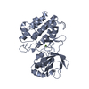
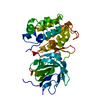
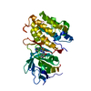
Method to determine structure: MOLECULAR REPLACEMENT Starting model: apo murine NIK kinase domain Resolution: 2.5→47.73 Å / Cor.coef. Fo:Fc: 0.948 / Cor.coef. Fo:Fc free: 0.913 / SU B: 17.602 / SU ML: 0.19 / Cross valid method: THROUGHOUT / ESU R: 0.381 / ESU R Free: 0.262 / Stereochemistry target values: MAXIMUM LIKELIHOOD / Details: HYDROGENS HAVE BEEN USED IF PRESENT IN THE INPUT
Rfactor
Num. reflection
% reflection
Selection details
Rfree
0.24047
3259
10.1 %
RANDOM
Rwork
0.18931
-
-
-
all
0.19446
32110
-
-
obs
0.19446
32110
99.54 %
-
Solvent computation
Ion probe radii: 0.8 Å / Shrinkage radii: 0.8 Å / VDW probe radii: 1.4 Å / Solvent model: MASK
Displacement parameters
Biso mean: 45.801 Å2
Baniso -1
Baniso -2
Baniso -3
1-
-0.28 Å2
0 Å2
-0 Å2
2-
-
-0.28 Å2
0 Å2
3-
-
-
0.57 Å2
Refinement step
Cycle: LAST / Resolution: 2.5→47.73 Å
Protein
Nucleic acid
Ligand
Solvent
Total
Num. atoms
4956
0
69
61
5086
Refine LS restraints
Refine-ID
Type
Dev ideal
Dev ideal target
Number
X-RAY DIFFRACTION
r_bond_refined_d
0.007
0.019
5150
X-RAY DIFFRACTION
r_angle_refined_deg
1.224
1.992
6969
X-RAY DIFFRACTION
r_dihedral_angle_1_deg
5.697
5
634
X-RAY DIFFRACTION
r_dihedral_angle_2_deg
38.37
23.443
212
X-RAY DIFFRACTION
r_dihedral_angle_3_deg
16.331
15
892
X-RAY DIFFRACTION
r_dihedral_angle_4_deg
18.466
15
37
X-RAY DIFFRACTION
r_chiral_restr
0.08
0.2
748
X-RAY DIFFRACTION
r_gen_planes_refined
0.005
0.021
3858
LS refinement shell
Resolution: 2.5→2.552 Å / Total num. of bins used: 25
Rfactor
Num. reflection
% reflection
Rfree
0.365
163
-
Rwork
0.295
1574
-
obs
-
-
99.14 %
Refinement TLS params.
Method: refined / Refine-ID: X-RAY DIFFRACTION
ID
L11 (°2)
L12 (°2)
L13 (°2)
L22 (°2)
L23 (°2)
L33 (°2)
S11 (Å °)
S12 (Å °)
S13 (Å °)
S21 (Å °)
S22 (Å °)
S23 (Å °)
S31 (Å °)
S32 (Å °)
S33 (Å °)
T11 (Å2)
T12 (Å2)
T13 (Å2)
T22 (Å2)
T23 (Å2)
T33 (Å2)
Origin x (Å)
Origin y (Å)
Origin z (Å)
1
3.0346
-0.7101
-1.6015
5.4137
0.1059
1.6367
0.067
0.0915
0.2536
-0.0639
-0.0858
-0.2112
0.0415
-0.0361
0.0188
0.0576
0.0679
0.0136
0.0915
0.0208
0.2102
46.3999
40.0639
-3.037
2
2.7396
0.4319
-0.1751
4.3899
-0.3596
3.9335
-0.0918
0.1618
-0.1261
-0.2607
0.0187
-0.2023
0.1244
0.2347
0.0731
0.1887
0.056
0.0378
0.0426
0.0031
0.017
53.0599
12.4309
-4.2842
3
3.5853
1.3026
-1.1479
5.4904
0.67
2.3037
0.1
0.0162
0.0281
0.0406
-0.0803
-0.2389
-0.0432
0.0091
-0.0197
0.0089
0.0202
-0.0229
0.0927
-0.03
0.18
31.3036
67.0408
2.6789
4
3.4109
0.1693
0.5093
4.1307
0.4486
4.7559
0.2121
0.0146
-0.172
0.0469
-0.0633
-0.0196
0.4816
0.0103
-0.1488
0.081
-0.0391
-0.029
0.1124
0.0435
0.0546
17.3244
42.9371
1.0622
Refinement TLS group
ID
Refine-ID
Refine TLS-ID
Auth asym-ID
Auth seq-ID
1
X-RAY DIFFRACTION
1
A
334 - 472
2
X-RAY DIFFRACTION
2
A
473 - 671
3
X-RAY DIFFRACTION
3
B
334 - 472
4
X-RAY DIFFRACTION
4
B
473 - 674
+
About Yorodumi
-
News
-
Feb 9, 2022. New format data for meta-information of EMDB entries
New format data for meta-information of EMDB entries
Version 3 of the EMDB header file is now the official format.
The previous official version 1.9 will be removed from the archive.
In the structure databanks used in Yorodumi, some data are registered as the other names, "COVID-19 virus" and "2019-nCoV". Here are the details of the virus and the list of structure data.
Jan 31, 2019. EMDB accession codes are about to change! (news from PDBe EMDB page)
EMDB accession codes are about to change! (news from PDBe EMDB page)
The allocation of 4 digits for EMDB accession codes will soon come to an end. Whilst these codes will remain in use, new EMDB accession codes will include an additional digit and will expand incrementally as the available range of codes is exhausted. The current 4-digit format prefixed with “EMD-” (i.e. EMD-XXXX) will advance to a 5-digit format (i.e. EMD-XXXXX), and so on. It is currently estimated that the 4-digit codes will be depleted around Spring 2019, at which point the 5-digit format will come into force.
The EM Navigator/Yorodumi systems omit the EMD- prefix.
Related info.:Q: What is EMD? / ID/Accession-code notation in Yorodumi/EM Navigator
Yorodumi is a browser for structure data from EMDB, PDB, SASBDB, etc.
This page is also the successor to EM Navigator detail page, and also detail information page/front-end page for Omokage search.
The word "yorodu" (or yorozu) is an old Japanese word meaning "ten thousand". "mi" (miru) is to see.
Related info.:EMDB / PDB / SASBDB / Comparison of 3 databanks / Yorodumi Search / Aug 31, 2016. New EM Navigator & Yorodumi / Yorodumi Papers / Jmol/JSmol / Function and homology information / Changes in new EM Navigator and Yorodumi
 Movie
Movie Controller

 Yorodumi
 Open data
 Basic information
 Components
 Keywords
 Function and homology information
 X-RAY DIFFRACTION /
  Authors
 Citation
 Structure visualization
 Downloads & links
 Other downloads
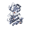


 PDBj
PDBj
 Assembly


 Spodoptera frugiperda (fall armyworm) / Strain (production host): SF9
 Mass: 96.063 Da / Num. of mol.: 3 / Source method: obtained synthetically / Formula: SO4
 Mass: 397.881 Da / Num. of mol.: 2 / Source method: obtained synthetically / Formula: C19H16ClN5OS
 Mass: 18.015 Da / Num. of mol.: 61 / Source method: isolated from a natural source / Formula: H2O
 Sample preparation
 / Beamline: BL7-1 / Wavelength: 0.97946 Å
 Processing