 Movie
Movie Controller


+ Open data

- Basic information
| Entry | Database: PDB / ID: 6mtm | ||||||
|---|---|---|---|---|---|---|---|
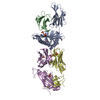
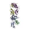
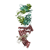
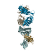
| Title | Crystal Structure of EM2 TCR in complex with HLA-B*37:01-NP338 | ||||||
 Components Components |
| ||||||
 Keywords Keywords | IMMUNE SYSTEM / TCR / T cell / influenza / HLA / HLA-B18 / HLA-B37 / HLA-B44 / viral mutation / CD8 T cells | ||||||
| Function / homology |  Function and homology information Function and homology informationregulation of interleukin-12 production / regulation of dendritic cell differentiation / regulation of T cell anergy / regulation of interleukin-6 production / protection from natural killer cell mediated cytotoxicity / TAP binding / detection of bacterium / antigen processing and presentation of endogenous peptide antigen via MHC class Ib / antigen processing and presentation of endogenous peptide antigen via MHC class I via ER pathway, TAP-independent / early endosome lumen ...regulation of interleukin-12 production / regulation of dendritic cell differentiation / regulation of T cell anergy / regulation of interleukin-6 production / protection from natural killer cell mediated cytotoxicity / TAP binding / detection of bacterium / antigen processing and presentation of endogenous peptide antigen via MHC class Ib / antigen processing and presentation of endogenous peptide antigen via MHC class I via ER pathway, TAP-independent / early endosome lumen / Nef mediated downregulation of MHC class I complex cell surface expression / DAP12 interactions / secretory granule membrane / Endosomal/Vacuolar pathway / T cell mediated cytotoxicity / Antigen Presentation: Folding, assembly and peptide loading of class I MHC / lumenal side of endoplasmic reticulum membrane / regulation of iron ion transport / cellular response to iron(III) ion / negative regulation of iron ion transport / negative regulation of forebrain neuron differentiation / antigen processing and presentation of exogenous protein antigen via MHC class Ib, TAP-dependent / peptide antigen assembly with MHC class I protein complex / regulation of erythrocyte differentiation / ER to Golgi transport vesicle membrane / response to molecule of bacterial origin / defense response / HFE-transferrin receptor complex / MHC class I peptide loading complex / transferrin transport / cellular response to iron ion / negative regulation of receptor-mediated endocytosis / positive regulation of T cell cytokine production / antigen processing and presentation of endogenous peptide antigen via MHC class I / MHC class I protein complex / peptide antigen assembly with MHC class II protein complex / negative regulation of neurogenesis / cellular response to nicotine / MHC class II protein complex / positive regulation of receptor-mediated endocytosis / multicellular organismal-level iron ion homeostasis / positive regulation of T cell mediated cytotoxicity / specific granule lumen / antigen processing and presentation of exogenous peptide antigen via MHC class II / positive regulation of immune response / peptide antigen binding / phagocytic vesicle membrane / recycling endosome membrane / negative regulation of epithelial cell proliferation / positive regulation of T cell activation / Interferon gamma signaling / Immunoregulatory interactions between a Lymphoid and a non-Lymphoid cell / Interferon alpha/beta signaling / Modulation by Mtb of host immune system / sensory perception of smell / tertiary granule lumen / positive regulation of cellular senescence / MHC class II protein complex binding / T cell differentiation in thymus / DAP12 signaling / late endosome membrane / negative regulation of neuron projection development / protein refolding / protein-folding chaperone binding / ER-Phagosome pathway / early endosome membrane / amyloid fibril formation / protein homotetramerization / intracellular iron ion homeostasis / adaptive immune response / learning or memory / immune response / endoplasmic reticulum lumen / Amyloid fiber formation / signaling receptor binding / Golgi membrane / external side of plasma membrane / innate immune response / lysosomal membrane / focal adhesion / Neutrophil degranulation / SARS-CoV-2 activates/modulates innate and adaptive immune responses / structural molecule activity / cell surface / Golgi apparatus / endoplasmic reticulum / protein homodimerization activity / : / extracellular exosome / extracellular region / membrane / identical protein binding / plasma membrane / cytosol Similarity search - Function | ||||||
| Biological species |  Homo sapiens (human) Homo sapiens (human)  unidentified influenza virus unidentified influenza virus | ||||||
| Method |  X-RAY DIFFRACTION / SYNCHROTRON / MOLECULAR REPLACEMENT / molecular replacement / Resolution: 3 Å X-RAY DIFFRACTION / SYNCHROTRON / MOLECULAR REPLACEMENT / molecular replacement / Resolution: 3 Å | ||||||
 Authors Authors | Gras, S. | ||||||
 Citation Citation | Journal: Nat Commun / Year: 2018 Title: Broad CD8+T cell cross-recognition of distinct influenza A strains in humans. Authors: Grant, E.J. / Josephs, T.M. / Loh, L. / Clemens, E.B. / Sant, S. / Bharadwaj, M. / Chen, W. / Rossjohn, J. / Gras, S. / Kedzierska, K. | ||||||
| History |
|
- Structure visualization
| Structure viewer | Molecule: MolmilJmol/JSmol |
|---|
- Downloads & links
-Download
| PDBx/mmCIF format | 6mtm.cif.gz | 175.5 KB | Display | PDBx/mmCIF format |
|---|---|---|---|---|
| PDB format | pdb6mtm.ent.gz | 136.9 KB | Display | PDB format |
| PDBx/mmJSON format | 6mtm.json.gz | Tree view | PDBx/mmJSON format | |
| Others |  Other downloads Other downloads |
-Validation report
| Arichive directory | https://data.pdbj.org/pub/pdb/validation_reports/mt/6mtmftp://data.pdbj.org/pub/pdb/validation_reports/mt/6mtm | HTTPS FTP |
|---|
-Related structure data
| Related structure data |  6mt3C  6mt4C  6mt5C  6mt6C  6mtlC  1kgcS  3gsoS S: Starting model for refinement C: citing same article ( |
|---|---|
| Similar structure data |


-Links
 PDBj
PDBj

- Assembly
| Deposited unit | 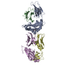
| ||||||||
|---|---|---|---|---|---|---|---|---|---|
| 1 |
| ||||||||
| Unit cell |
|
-Components
| #1: Protein | Mass: 31927.051 Da / Num. of mol.: 1 Source method: isolated from a genetically manipulated source Source: (gene. exp.) Homo sapiens (human) / Gene: HLA-B, HLAB / Plasmid: pET30 / Production host:  |
|---|---|
| #2: Protein | Mass: 11748.160 Da / Num. of mol.: 1 Source method: isolated from a genetically manipulated source Source: (gene. exp.) Homo sapiens (human) / Gene: B2M, CDABP0092, HDCMA22P / Plasmid: pET30 / Production host: |
| #3: Protein/peptide | Mass: 1126.282 Da / Num. of mol.: 1 / Source method: obtained synthetically / Source: (synth.) unidentified influenza virus |
| #4: Protein | Mass: 21488.000 Da / Num. of mol.: 1 Source method: isolated from a genetically manipulated source Source: (gene. exp.) Homo sapiens (human) / Plasmid: pET30 / Production host: |
| #5: Protein | Mass: 26891.947 Da / Num. of mol.: 1 Source method: isolated from a genetically manipulated source Source: (gene. exp.) Homo sapiens (human) / Plasmid: pET30 / Production host: |
| Has protein modification | Y |
-Experimental details
-Experiment
| Experiment | Method: X-RAY DIFFRACTION / Number of used crystals: 1 |
|---|
- Sample preparation
| Crystal | Density Matthews: 2.33 Å3/Da / Density % sol: 47.24 % |
|---|---|
| Crystal grow | Temperature: 277 K / Method: vapor diffusion, hanging drop / pH: 8 / Details: 19% PEG 3350, 0.2 ammonium tartrate |
-Data collection
| Diffraction | Mean temperature: 100 K / Serial crystal experiment: N |
|---|---|
| Diffraction source | Source: SYNCHROTRON / Site: Australian Synchrotron  / Beamline: MX2 / Wavelength: 0.954 Å / Beamline: MX2 / Wavelength: 0.954 Å |
| Detector | Type: ADSC QUANTUM 315r / Detector: CCD / Date: Oct 22, 2014 |
| Radiation | Protocol: SINGLE WAVELENGTH / Monochromatic (M) / Laue (L): M / Scattering type: x-ray |
| Radiation wavelength | Wavelength: 0.954 Å / Relative weight: 1 |
| Reflection | Resolution: 3→47.4 Å / Num. obs: 18278 / % possible obs: 100 % / Redundancy: 7.2 % / Biso Wilson estimate: 39.91 Å2 / Rpim(I) all: 0.134 / Net I/σ(I): 6.3 |
| Reflection shell | Resolution: 3→3.16 Å / Num. unique obs: 2614 / Rpim(I) all: 0.391 |
-Phasing
| Phasing | Method: molecular replacement |
|---|
- Processing
| Software |
| ||||||||||||||||||||||||||||||||||||||||||||||||||||||||||||||||||||||||||||||||||||||||||||||||||||||||||||
|---|---|---|---|---|---|---|---|---|---|---|---|---|---|---|---|---|---|---|---|---|---|---|---|---|---|---|---|---|---|---|---|---|---|---|---|---|---|---|---|---|---|---|---|---|---|---|---|---|---|---|---|---|---|---|---|---|---|---|---|---|---|---|---|---|---|---|---|---|---|---|---|---|---|---|---|---|---|---|---|---|---|---|---|---|---|---|---|---|---|---|---|---|---|---|---|---|---|---|---|---|---|---|---|---|---|---|---|---|---|
| Refinement | Method to determine structure: MOLECULAR REPLACEMENT Starting model: 3GSO, 1kgc Resolution: 3→45.44 Å / Cor.coef. Fo:Fc: 0.835 / Cor.coef. Fo:Fc free: 0.701 / Cross valid method: THROUGHOUT / σ(F): 0 / SU Rfree Blow DPI: 0.533
| ||||||||||||||||||||||||||||||||||||||||||||||||||||||||||||||||||||||||||||||||||||||||||||||||||||||||||||
| Displacement parameters | Biso max: 110.53 Å2 / Biso mean: 27.35 Å2 / Biso min: 3 Å2
| ||||||||||||||||||||||||||||||||||||||||||||||||||||||||||||||||||||||||||||||||||||||||||||||||||||||||||||
| Refine analyze | Luzzati coordinate error obs: 0.44 Å | ||||||||||||||||||||||||||||||||||||||||||||||||||||||||||||||||||||||||||||||||||||||||||||||||||||||||||||
| Refinement step | Cycle: final / Resolution: 3→45.44 Å
| ||||||||||||||||||||||||||||||||||||||||||||||||||||||||||||||||||||||||||||||||||||||||||||||||||||||||||||
| Refine LS restraints |
| ||||||||||||||||||||||||||||||||||||||||||||||||||||||||||||||||||||||||||||||||||||||||||||||||||||||||||||
| LS refinement shell | Resolution: 3→3.18 Å / Rfactor Rfree error: 0 / Total num. of bins used: 9
|
