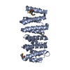
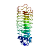
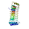
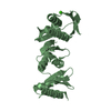
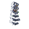
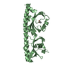
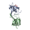
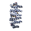
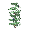
[日本語] English
- PDB-6mq5: Structure of human CLASP1 TOG1 -

+
データを開く


IDまたはキーワード:

読み込み中...

-
基本情報

登録情報
データベース: PDB / ID: 6mq5
タイトルStructure of human CLASP1 TOG1
要素CLIP-associating protein 1
キーワードSTRUCTURAL PROTEIN / Microtubule Plus End Binding
機能・相同性
機能・相同性情報


centrosomal corona / establishment of spindle orientation / positive regulation of basement membrane assembly involved in embryonic body morphogenesis / negative regulation of microtubule polymerization or depolymerization / negative regulation of wound healing, spreading of epidermal cells / regulation of gastrulation / vesicle targeting / microtubule anchoring / cortical microtubule cytoskeleton / basal cortex ...centrosomal corona / establishment of spindle orientation / positive regulation of basement membrane assembly involved in embryonic body morphogenesis / negative regulation of microtubule polymerization or depolymerization / negative regulation of wound healing, spreading of epidermal cells / regulation of gastrulation / vesicle targeting / microtubule anchoring / cortical microtubule cytoskeleton / basal cortex / positive regulation of extracellular matrix disassembly / microtubule organizing center organization / kinetochore microtubule / establishment of mitotic spindle localization / astral microtubule organization / Role of ABL in ROBO-SLIT signaling / kinetochore binding / regulation of epithelial to mesenchymal transition / establishment of epithelial cell polarity / dystroglycan binding / microtubule plus-end / microtubule nucleation / negative regulation of microtubule depolymerization / microtubule bundle formation / microtubule plus-end binding / exit from mitosis / negative regulation of stress fiber assembly / microtubule organizing center / regulation of focal adhesion assembly / establishment or maintenance of cell polarity / positive regulation of epithelial cell migration / positive regulation of exocytosis / Golgi organization / cytoplasmic microtubule / mitotic spindle assembly / Amplification of signal from unattached kinetochores via a MAD2 inhibitory signal / regulation of microtubule cytoskeleton organization / Mitotic Prometaphase / EML4 and NUDC in mitotic spindle formation / Loss of Nlp from mitotic centrosomes / Loss of proteins required for interphase microtubule organization from the centrosome / Recruitment of mitotic centrosome proteins and complexes / positive regulation of microtubule polymerization / Recruitment of NuMA to mitotic centrosomes / Resolution of Sister Chromatid Cohesion / Anchoring of the basal body to the plasma membrane / AURKA Activation by TPX2 / mitotic spindle organization / RHO GTPases Activate Formins / spindle microtubule / mitotic spindle / kinetochore / microtubule cytoskeleton organization / Separation of Sister Chromatids / Regulation of PLK1 Activity at G2/M Transition / cell cortex / microtubule binding / cell division / centrosome / Golgi apparatus / membrane / cytosol
類似検索 - 分子機能
: / XMAP215/Dis1/CLASP, TOG domain / CLASP N-terminal domain / CLASP N terminal / Centrosomal protein CEP104-like, TOG domain / TOG domain / TOG / HEAT repeat / HEAT repeat profile. / HEAT, type 2 ...: / XMAP215/Dis1/CLASP, TOG domain / CLASP N-terminal domain / CLASP N terminal / Centrosomal protein CEP104-like, TOG domain / TOG domain / TOG / HEAT repeat / HEAT repeat profile. / HEAT, type 2 / Armadillo-like helical / Armadillo-type fold
類似検索 - ドメイン・相同性
CLIP-associating protein 1
類似検索 - 構成要素
生物種Homo sapiens (ヒト)
手法X線回折 / シンクロトロン / 単波長異常分散 / 解像度: 2.146 Å
データ登録者Leano, J.B. / Slep, K.C.
資金援助 米国, 1件
組織認可番号
National Institutes of Health/National Institute of General Medical Sciences (NIH/NIGMS)R01GM094415 米国
引用ジャーナル: Plos One / : 2019
タイトル: Structures of TOG1 and TOG2 from the human microtubule dynamics regulator CLASP1.
著者: Leano, J.B. / Slep, K.C.
履歴
登録2018年10月9日登録サイト: RCSB / 処理サイト: RCSB
改定 1.02019年7月17日Provider: repository / タイプ: Initial release
改定 1.12019年7月31日Group: Data collection / Database references / カテゴリ: citation / citation_author
Item: _citation.country / _citation.journal_abbrev ..._citation.country / _citation.journal_abbrev / _citation.journal_id_CSD / _citation.journal_id_ISSN / _citation.journal_volume / _citation.page_first / _citation.page_last / _citation.pdbx_database_id_DOI / _citation.pdbx_database_id_PubMed / _citation.title / _citation.year / _citation_author.identifier_ORCID
改定 1.22020年1月1日Group: Author supporting evidence / カテゴリ: pdbx_audit_support / Item: _pdbx_audit_support.funding_organization

-
構造の表示

構造ビューア分子:
MolmilJmol/JSmol

ダウンロードとリンク

-
集合体

登録構造単位
A: CLIP-associating protein 1
B: CLIP-associating protein 1


分子量 (理論値)分子数
合計 (水以外)58,0322
ポリマ-58,0322
非ポリマー00
3,297183
1
A: CLIP-associating protein 1


分子量 (理論値)分子数
合計 (水以外)29,0161
ポリマ-29,0161
非ポリマー00
181
タイプ名称対称操作
identity operation1_555x,y,z1
手法PISA
2
B: CLIP-associating protein 1


分子量 (理論値)分子数
合計 (水以外)29,0161
ポリマ-29,0161
非ポリマー00
181
タイプ名称対称操作
identity operation1_555x,y,z1
手法PISA
単位格子
Length a, b, c (Å)40.975, 114.022, 46.649
Angle α, β, γ (deg.)90.00, 99.85, 90.00
Int Tables number4
Space group name H-MP1211

-
要素

#1: タンパク質 CLIP-associating protein 1 / Cytoplasmic linker-associated protein 1 / Multiple asters homolog 1 / Protein Orbit homolog 1 / hOrbit1


分子量: 29015.918 Da / 分子数: 2 / 由来タイプ: 組換発現 / 由来: (組換発現) Homo sapiens (ヒト) / 遺伝子: CLASP1, KIAA0622, MAST1 / 発現宿主: Escherichia coli (大腸菌) / 参照: UniProt: Q7Z460
#2: 水 ChemComp-HOH / water


分子量: 18.015 Da / 分子数: 183 / 由来タイプ: 天然 / : H2O

-
実験情報

-
実験

実験手法: X線回折 / 使用した結晶の数: 1

-
試料調製

結晶マシュー密度: 2.01 Å3/Da / 溶媒含有率: 33.52 %
結晶化温度: 291 K / 手法: 蒸気拡散法, ハンギングドロップ法 / pH: 6.5
詳細: equal volume of 10 mg/ml hCLASP1 TOG1 and well solution containing 30% PEG600, 100mM MES, pH 6.5, 10% glycerol were mixed together and equilibrated over 1 ml of well solution, crystals that ...詳細: equal volume of 10 mg/ml hCLASP1 TOG1 and well solution containing 30% PEG600, 100mM MES, pH 6.5, 10% glycerol were mixed together and equilibrated over 1 ml of well solution, crystals that were originally crystallized in 1.5 M sodium malonate, pH 6.25, 18C, were used for microseeding

-
データ収集

回折平均測定温度: 100 K / Serial crystal experiment: N
放射光源由来: シンクロトロン / サイト: APS / ビームライン: 22-ID / 波長: 0.97964 Å
検出器タイプ: MAR scanner 300 mm plate / 検出器: IMAGE PLATE / 日付: 2014年8月6日
放射プロトコル: SINGLE WAVELENGTH / 単色(M)・ラウエ(L): M / 散乱光タイプ: x-ray
放射波長波長: 0.97964 Å / 相対比: 1
反射解像度: 2.146→50 Å / Num. obs: 41883 / % possible obs: 92.3 % / 冗長度: 3.5 % / CC1/2: 1 / Rpim(I) all: 0.08 / Rrim(I) all: 0.155 / Net I/σ(I): 9.6
反射 シェル解像度: 2.15→2.23 Å / 冗長度: 3 % / Mean I/σ(I) obs: 1.96 / Num. unique obs: 3667 / CC1/2: 0.82 / Rpim(I) all: 0.348 / Rrim(I) all: 0.646 / % possible all: 81.2

-
解析

ソフトウェア
名称バージョン分類
PHENIX(1.14_3211: ???)精密化
HKL-2000データ削減
HKL-2000データスケーリング
AutoSol位相決定
精密化構造決定の手法: 単波長異常分散 / 解像度: 2.146→32.947 Å / SU ML: 0.27 / 交差検証法: FREE R-VALUE / σ(F): 0 / 位相誤差: 25.97
Rfactor反射数%反射
Rfree0.2437 1931 9.18 %
Rwork0.1899 --
obs0.1949 21035 91.16 %
溶媒の処理減衰半径: 0.9 Å / VDWプローブ半径: 1.11 Å
精密化ステップサイクル: LAST / 解像度: 2.146→32.947 Å
タンパク質核酸リガンド溶媒全体
原子数3676 0 0 183 3859
拘束条件
Refine-IDタイプDev ideal
X-RAY DIFFRACTIONf_bond_d0.0113732
X-RAY DIFFRACTIONf_angle_d1.3815030
X-RAY DIFFRACTIONf_dihedral_angle_d16.0551434
X-RAY DIFFRACTIONf_chiral_restr0.223585
X-RAY DIFFRACTIONf_plane_restr0.006650
LS精密化 シェル
解像度 (Å)Rfactor RfreeNum. reflection RfreeRfactor RworkNum. reflection RworkRefine-ID% reflection obs (%)
2.1459-2.19960.34341130.24791129X-RAY DIFFRACTION76
2.1996-2.2590.29361190.23251200X-RAY DIFFRACTION80
2.259-2.32550.2851310.21391303X-RAY DIFFRACTION86
2.3255-2.40050.28621290.20391274X-RAY DIFFRACTION86
2.4005-2.48630.22091300.19841302X-RAY DIFFRACTION88
2.4863-2.58580.26171320.19521335X-RAY DIFFRACTION88
2.5858-2.70350.29121360.21271352X-RAY DIFFRACTION91
2.7035-2.84590.30251390.22791369X-RAY DIFFRACTION93
2.8459-3.02410.28491460.21911418X-RAY DIFFRACTION95
3.0241-3.25740.28221470.20611461X-RAY DIFFRACTION97
3.2574-3.58490.24111510.18061471X-RAY DIFFRACTION98
3.5849-4.10280.20411520.16161487X-RAY DIFFRACTION99
4.1028-5.16580.20061520.15951499X-RAY DIFFRACTION99
5.1658-32.95090.19181540.171504X-RAY DIFFRACTION100

+
万見について

-
お知らせ

-
2022年2月9日: EMDBエントリの付随情報ファイルのフォーマットが新しくなりました

EMDBエントリの付随情報ファイルのフォーマットが新しくなりました

  • EMDBのヘッダファイルのバージョン3が、公式のフォーマットとなりました。
  • これまでは公式だったバージョン1.9は、アーカイブから削除されます。

関連情報:EMDBヘッダ

外部リンク:wwPDBはEMDBデータモデルのバージョン3へ移行します

-
2020年8月12日: 新型コロナ情報

新型コロナ情報

URL: https://pdbjlvh1.pdbj.org/emnavi/covid19.php

新ページ: EM Navigatorに新型コロナウイルスの特設ページを開設しました。

関連情報:Covid-19情報 / 2020年3月5日: 新型コロナウイルスの構造データ

+
2020年3月5日: 新型コロナウイルスの構造データ

新型コロナウイルスの構造データ

関連情報:万見生物種 / 2020年8月12日: 新型コロナ情報

外部リンク:COVID-19特集ページ - PDBj / 今月の分子2020年2月:コロナウイルスプロテーアーゼ

+
2019年1月31日: EMDBのIDの桁数の変更

EMDBのIDの桁数の変更

  • EMDBエントリに付与されているアクセスコード(EMDB-ID)は4桁の数字(例、EMD-1234)でしたが、間もなく枯渇します。これまでの4桁のID番号は4桁のまま変更されませんが、4桁の数字を使い切った後に発行されるIDは5桁以上の数字(例、EMD-12345)になります。5桁のIDは2019年の春頃から発行される見通しです。
  • EM Navigator/万見では、接頭語「EMD-」は省略されています。

関連情報:Q: 「EMD」とは何ですか? / 万見/EM NavigatorにおけるID/アクセスコードの表記

外部リンク:EMDB Accession Codes are Changing Soon! / PDBjへお問い合わせ

+
2017年7月12日: PDB大規模アップデート

PDB大規模アップデート

  • 新バージョンのPDBx/mmCIF辞書形式に基づくデータがリリースされました。
  • 今回の更新はバージョン番号が4から5になる大規模なもので、全エントリデータの書き換えが行われる「Remediation」というアップデートに該当します。
  • このバージョンアップで、電子顕微鏡の実験手法に関する多くの項目の書式が改定されました(例:em_softwareなど)。
  • EM NavigatorとYorodumiでも、この改定に基づいた表示内容になります。

外部リンク:wwPDB Remediation / OneDepデータ基準に準拠した、より強化された内容のモデル構造ファイルが、PDBアーカイブで公開されました。

-
万見 (Yorodumi)

幾万の構造データを、幾万の視点から

  • 万見(Yorodumi)は、EMDB/PDB/SASBDBなどの構造データを閲覧するためのページです。
  • EM Navigatorの詳細ページの後継、Omokage検索のフロントエンドも兼ねています。

関連情報:EMDB / PDB / SASBDB / 3つのデータバンクの比較 / 万見検索 / 2016年8月31日: 新しいEM Navigatorと万見 / 万見文献 / Jmol/JSmol / 機能・相同性情報 / 新しいEM Navigatorと万見の変更点

他の情報も見る