 Movie
Movie Controller

[English] 日本語
 Yorodumi
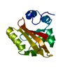
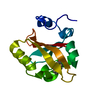
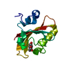
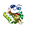
- PDB-6mkt: Photoactive Yellow Protein with 3-chlorotyrosine substituted at p... -
+ Open data

- Basic information
| Entry | Database: PDB / ID: 6mkt | |||||||||
|---|---|---|---|---|---|---|---|---|---|---|
| Title | Photoactive Yellow Protein with 3-chlorotyrosine substituted at position 42 | |||||||||
 Components Components | Photoactive yellow protein | |||||||||
 Keywords Keywords | SIGNALING PROTEIN / chlorinated chromophore / hydrogen bonding network / active site / low-barrier hydrogen bond | |||||||||
| Function / homology |  Function and homology information Function and homology informationphotoreceptor activity / phototransduction / regulation of DNA-templated transcription / identical protein binding Similarity search - Function | |||||||||
| Biological species |  Halorhodospira halophila (bacteria) Halorhodospira halophila (bacteria) | |||||||||
| Method |  X-RAY DIFFRACTION / SYNCHROTRON / MOLECULAR REPLACEMENT / Resolution: 1.6 Å X-RAY DIFFRACTION / SYNCHROTRON / MOLECULAR REPLACEMENT / Resolution: 1.6 Å | |||||||||
 Authors Authors | Thomson, B.D. / Both, J. / Wu, Y. / Parrish, R.M. / Martinez, T. / Boxer, S.G. | |||||||||
| Funding support |  United States, 2items United States, 2items
| |||||||||
 Citation Citation | Journal: J.Phys.Chem.B / Year: 2019 Title: Perturbation of Short Hydrogen Bonds in Photoactive Yellow Protein via Noncanonical Amino Acid Incorporation. Authors: Thomson, B. / Both, J. / Wu, Y. / Parrish, R.M. / Martinez, T.J. / Boxer, S.G. | |||||||||
| History |
|
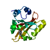
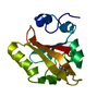
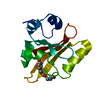
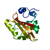
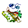
- Structure visualization
| Structure viewer | Molecule: MolmilJmol/JSmol |
|---|
- Downloads & links
-Download
| PDBx/mmCIF format | 6mkt.cif.gz | 42.6 KB | Display | PDBx/mmCIF format |
|---|---|---|---|---|
| PDB format | pdb6mkt.ent.gz | 27 KB | Display | PDB format |
| PDBx/mmJSON format | 6mkt.json.gz | Tree view | PDBx/mmJSON format | |
| Others |  Other downloads Other downloads |
-Validation report
| Arichive directory | https://data.pdbj.org/pub/pdb/validation_reports/mk/6mktftp://data.pdbj.org/pub/pdb/validation_reports/mk/6mkt | HTTPS FTP |
|---|
-Related structure data
| Related structure data |  6mhiC  6mhnC  6mmdC  1nwzS S: Starting model for refinement C: citing same article ( |
|---|---|
| Similar structure data |

-Links
 PDBj
PDBj


- Assembly
| Deposited unit | 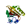
| ||||||||
|---|---|---|---|---|---|---|---|---|---|
| 1 |
| ||||||||
| Unit cell |
|
-Components
| #1: Protein | Mass: 13923.021 Da / Num. of mol.: 1 Source method: isolated from a genetically manipulated source Source: (gene. exp.) Halorhodospira halophila (bacteria) / Gene: pyp / Production host: |
|---|---|
| #2: Chemical | ChemComp-HC4 /   Mass: 164.158 Da / Num. of mol.: 1 / Source method: obtained synthetically / Formula: C9H8O3 Mass: 164.158 Da / Num. of mol.: 1 / Source method: obtained synthetically / Formula: C9H8O3 |
| #3: Water | ChemComp-HOH /  Mass: 18.015 Da / Num. of mol.: 92 / Source method: isolated from a natural source / Formula: H2O Mass: 18.015 Da / Num. of mol.: 92 / Source method: isolated from a natural source / Formula: H2O |
-Experimental details
-Experiment
| Experiment | Method: X-RAY DIFFRACTION / Number of used crystals: 1 |
|---|
- Sample preparation
| Crystal | Density Matthews: 1.75 Å3/Da / Density % sol: 29.58 % |
|---|---|
| Crystal grow | Temperature: 298 K / Method: vapor diffusion, hanging drop Details: 1M sodium chloride, 2.4M ammonium sulfate, pH = 6.0, drops were microseeded |
-Data collection
| Diffraction | Mean temperature: 80 K |
|---|---|
| Diffraction source | Source: SYNCHROTRON / Site: SSRL / Beamline: BL14-1 / Wavelength: 1.18076 Å |
| Detector | Type: RAYONIX MX325HE / Detector: CCD / Date: Nov 20, 2015 |
| Radiation | Protocol: SINGLE WAVELENGTH / Monochromatic (M) / Laue (L): M / Scattering type: x-ray |
| Radiation wavelength | Wavelength: 1.18076 Å / Relative weight: 1 |
| Reflection | Resolution: 1.21→50 Å / Num. obs: 13140 / % possible obs: 77.5 % / Redundancy: 9.9 % / Rmerge(I) obs: 0.075 / Net I/σ(I): 14 |
| Reflection shell | Resolution: 1.21→1.23 Å |
- Processing
| Software |
| ||||||||||||||||||||||||||||||||||||||||||
|---|---|---|---|---|---|---|---|---|---|---|---|---|---|---|---|---|---|---|---|---|---|---|---|---|---|---|---|---|---|---|---|---|---|---|---|---|---|---|---|---|---|---|---|
| Refinement | Method to determine structure: MOLECULAR REPLACEMENT Starting model: 1NWZ Resolution: 1.6→32.896 Å / SU ML: 0.12 / Cross valid method: FREE R-VALUE / σ(F): 1.36 / Phase error: 17.93
| ||||||||||||||||||||||||||||||||||||||||||
| Solvent computation | Shrinkage radii: 0.9 Å / VDW probe radii: 1.11 Å | ||||||||||||||||||||||||||||||||||||||||||
| Refinement step | Cycle: LAST / Resolution: 1.6→32.896 Å
| ||||||||||||||||||||||||||||||||||||||||||
| Refine LS restraints |
| ||||||||||||||||||||||||||||||||||||||||||
| LS refinement shell |
|
