[English] 日本語
Yorodumi
- PDB-5hdc: Femtosecond Structural Dynamics Drives the Trans/Cis Isomerizatio... -

+
Open data


ID or keywords:

Loading...

-
Basic information

Entry
Database: PDB / ID: 5hdc
TitleFemtosecond Structural Dynamics Drives the Trans/Cis Isomerization in Photoactive Yellow Protein: 100 fs to 400 fs Structure
ComponentsPhotoactive yellow protein
KeywordsSIGNALING PROTEIN / PHOTORECEPTOR CIS TRANS ISOMERIZATION / TRANS CIS ISOMERIZATION / FREE ELECTRON LASER
Function / homology
Function and homology information


photoreceptor activity / phototransduction / regulation of DNA-templated transcription / identical protein binding
Similarity search - Function
Photoactive yellow-protein / PAS domain / Beta-Lactamase / PAS fold / PAS fold / PAS domain / PAS repeat profile. / PAS domain / PAS domain superfamily / 2-Layer Sandwich / Alpha Beta
Similarity search - Domain/homology
Photoactive yellow protein
Similarity search - Component
Biological speciesHalorhodospira halophila (bacteria)
MethodX-RAY DIFFRACTION / FREE ELECTRON LASER / Resolution: 1.6 Å
AuthorsPande, K. / Tenboer, J. / Schmidt, M.
Funding support United States, 2items
OrganizationGrant numberCountry
National Science Foundation (NSF, United States)NSF-0952643 United States
National Science Foundation (NSF, United States)NSF-1231306 United States
CitationJournal: Science / Year: 2016
Title: Femtosecond structural dynamics drives the trans/cis isomerization in photoactive yellow protein.
Authors: Pande, K. / Hutchison, C.D. / Groenhof, G. / Aquila, A. / Robinson, J.S. / Tenboer, J. / Basu, S. / Boutet, S. / DePonte, D.P. / Liang, M. / White, T.A. / Zatsepin, N.A. / Yefanov, O. / ...Authors: Pande, K. / Hutchison, C.D. / Groenhof, G. / Aquila, A. / Robinson, J.S. / Tenboer, J. / Basu, S. / Boutet, S. / DePonte, D.P. / Liang, M. / White, T.A. / Zatsepin, N.A. / Yefanov, O. / Morozov, D. / Oberthuer, D. / Gati, C. / Subramanian, G. / James, D. / Zhao, Y. / Koralek, J. / Brayshaw, J. / Kupitz, C. / Conrad, C. / Roy-Chowdhury, S. / Coe, J.D. / Metz, M. / Xavier, P.L. / Grant, T.D. / Koglin, J.E. / Ketawala, G. / Fromme, R. / Srajer, V. / Henning, R. / Spence, J.C. / Ourmazd, A. / Schwander, P. / Weierstall, U. / Frank, M. / Fromme, P. / Barty, A. / Chapman, H.N. / Moffat, K. / van Thor, J.J. / Schmidt, M.
History
DepositionJan 5, 2016Deposition site: RCSB / Processing site: RCSB
Revision 1.0May 25, 2016Provider: repository / Type: Initial release
Revision 1.1Sep 13, 2017Group: Author supporting evidence / Derived calculations / Category: pdbx_audit_support / pdbx_struct_oper_list
Item: _pdbx_audit_support.funding_organization / _pdbx_struct_oper_list.symmetry_operation
Revision 1.2Feb 14, 2018Group: Data collection / Category: diffrn_source
Item: _diffrn_source.pdbx_synchrotron_beamline / _diffrn_source.pdbx_synchrotron_site
Revision 1.3Nov 27, 2019Group: Author supporting evidence / Category: pdbx_audit_support / Item: _pdbx_audit_support.funding_organization
Revision 1.4Aug 16, 2023Group: Data collection / Database references
Category: chem_comp_atom / chem_comp_bond ...chem_comp_atom / chem_comp_bond / database_2 / pdbx_related_exp_data_set
Item: _database_2.pdbx_DOI / _database_2.pdbx_database_accession
Revision 1.5Nov 15, 2023Group: Data collection / Derived calculations / Category: chem_comp_atom / chem_comp_bond / struct_conn
Item: _chem_comp_atom.atom_id / _chem_comp_bond.atom_id_1 ..._chem_comp_atom.atom_id / _chem_comp_bond.atom_id_1 / _chem_comp_bond.atom_id_2 / _struct_conn.pdbx_leaving_atom_flag

-
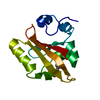
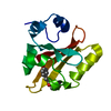
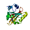
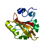
Structure visualization

Structure viewerMolecule:
MolmilJmol/JSmol

Downloads & links

-
Assembly

Deposited unit
A: Photoactive yellow protein


Theoretical massNumber of molelcules
Total (without water)14,0351
Polymers14,0351
Non-polymers00
Water1,65792
1


  • Idetical with deposited unit
  • defined by author
TypeNameSymmetry operationNumber
identity operation1_555x,y,z1
Unit cell
Length a, b, c (Å)66.900, 66.900, 40.800
Angle α, β, γ (deg.)90.00, 90.00, 120.00
Int Tables number173
Space group name H-MP63

-
Components

#1: Protein Photoactive yellow protein / / PYP


Mass: 14034.718 Da / Num. of mol.: 1
Source method: isolated from a genetically manipulated source
Source: (gene. exp.) Halorhodospira halophila (bacteria) / Gene: pyp / Production host: Escherichia coli (E. coli) / References: UniProt: P16113
#2: Water ChemComp-HOH / water / Water


Mass: 18.015 Da / Num. of mol.: 92 / Source method: isolated from a natural source / Formula: H2O

-
Experimental details

-
Experiment

ExperimentMethod: X-RAY DIFFRACTION / Number of used crystals: 1

-
Sample preparation

CrystalDensity Matthews: 1.88 Å3/Da / Density % sol: 34.5 %
Crystal growTemperature: 295 K / Method: liquid diffusion / pH: 7
Details: Combine and vortex 4.45M sodium malonate with 100 uL protein (135 mg/ml initial concentration) for a final sodium malonate concentration of 3.2M. Let stand at room temperature for 1 hour. ...Details: Combine and vortex 4.45M sodium malonate with 100 uL protein (135 mg/ml initial concentration) for a final sodium malonate concentration of 3.2M. Let stand at room temperature for 1 hour. Spin at 10,000g for 2 hours in small spin concentrators. Vortex and let stand at room temperature for 4 hours. Harvest crystals and rebuffer with 3.0M sodium malonate.
PH range: 6.95-7.05 / Temp details: Ambient Room Temperature

-
Data collection

DiffractionMean temperature: 295 K
Ambient temp details: Injection of crystals into vacuum. No temperature control.
Diffraction sourceSource: FREE ELECTRON LASER / Site: SLAC LCLS / Beamline: CXI / Wavelength: 1.31 Å
DetectorType: CS-PAD CXI-1 / Detector: PIXEL / Date: Mar 18, 2015
RadiationProtocol: SINGLE WAVELENGTH / Monochromatic (M) / Laue (L): M / Scattering type: x-ray
Radiation wavelengthWavelength: 1.31 Å / Relative weight: 1
ReflectionResolution: 1.6→19.31 Å / Num. all: 13039 / Num. obs: 13039 / % possible obs: 98.72 % / Redundancy: 300 % / Net I/σ(I): 10

-
Processing

Software
NameVersionClassification
REFMAC5.8.0069refinement
CrystFELdata reduction
CrystFELdata scaling
RefinementResolution: 1.6→19.31 Å / Cor.coef. Fo:Fc: 0.932 / Cor.coef. Fo:Fc free: 0.887 / SU B: 3.719 / SU ML: 0.119 / Cross valid method: THROUGHOUT / ESU R: 0.123 / ESU R Free: 0.123 / Stereochemistry target values: MAXIMUM LIKELIHOOD / Details: HYDROGENS HAVE BEEN ADDED IN THE RIDING POSITIONS
RfactorNum. reflection% reflectionSelection details
Rfree0.25832 655 4.8 %RANDOM
Rwork0.2083 ---
obs0.21068 13039 98.72 %-
Solvent computationIon probe radii: 0.8 Å / Shrinkage radii: 0.8 Å / VDW probe radii: 1.2 Å / Solvent model: MASK
Displacement parametersBiso mean: 5.462 Å2
Baniso -1Baniso -2Baniso -3
1--0.01 Å2-0.01 Å2-0 Å2
2---0.01 Å20 Å2
3---0.03 Å2
Refinement stepCycle: LAST / Resolution: 1.6→19.31 Å
ProteinNucleic acidLigandSolventTotal
Num. atoms987 0 0 92 1079
Refine LS restraints
Refine-IDTypeDev idealDev ideal targetNumber
X-RAY DIFFRACTIONr_bond_refined_d0.0220.0191044
X-RAY DIFFRACTIONr_bond_other_d0.0020.02969
X-RAY DIFFRACTIONr_angle_refined_deg2.3721.9411409
X-RAY DIFFRACTIONr_angle_other_deg2.5572.9932240
X-RAY DIFFRACTIONr_dihedral_angle_1_deg8.045124
X-RAY DIFFRACTIONr_dihedral_angle_2_deg39.6125.81855
X-RAY DIFFRACTIONr_dihedral_angle_3_deg21.615181
X-RAY DIFFRACTIONr_dihedral_angle_4_deg24.443153
X-RAY DIFFRACTIONr_chiral_restr0.1320.2150
X-RAY DIFFRACTIONr_gen_planes_refined0.0140.021198
X-RAY DIFFRACTIONr_gen_planes_other0.0020.02241
X-RAY DIFFRACTIONr_nbd_refined
X-RAY DIFFRACTIONr_nbd_other
X-RAY DIFFRACTIONr_nbtor_refined
X-RAY DIFFRACTIONr_nbtor_other
X-RAY DIFFRACTIONr_xyhbond_nbd_refined
X-RAY DIFFRACTIONr_xyhbond_nbd_other
X-RAY DIFFRACTIONr_metal_ion_refined
X-RAY DIFFRACTIONr_metal_ion_other
X-RAY DIFFRACTIONr_symmetry_vdw_refined
X-RAY DIFFRACTIONr_symmetry_vdw_other
X-RAY DIFFRACTIONr_symmetry_hbond_refined
X-RAY DIFFRACTIONr_symmetry_hbond_other
X-RAY DIFFRACTIONr_symmetry_metal_ion_refined
X-RAY DIFFRACTIONr_symmetry_metal_ion_other
X-RAY DIFFRACTIONr_mcbond_it0.4130.526499
X-RAY DIFFRACTIONr_mcbond_other0.4130.526498
X-RAY DIFFRACTIONr_mcangle_it0.7130.787622
X-RAY DIFFRACTIONr_mcangle_other0.7120.786623
X-RAY DIFFRACTIONr_scbond_it0.3130.51545
X-RAY DIFFRACTIONr_scbond_other0.3130.51545
X-RAY DIFFRACTIONr_scangle_it
X-RAY DIFFRACTIONr_scangle_other0.5090.761787
X-RAY DIFFRACTIONr_long_range_B_refined2.9934.2451222
X-RAY DIFFRACTIONr_long_range_B_other2.6514.1011202
X-RAY DIFFRACTIONr_rigid_bond_restr
X-RAY DIFFRACTIONr_sphericity_free
X-RAY DIFFRACTIONr_sphericity_bonded
LS refinement shellResolution: 1.6→1.641 Å / Total num. of bins used: 20
RfactorNum. reflection% reflection
Rfree0.368 38 -
Rwork0.305 897 -
obs--93.22 %

+
About Yorodumi

-
News

-
Feb 9, 2022. New format data for meta-information of EMDB entries

New format data for meta-information of EMDB entries

  • Version 3 of the EMDB header file is now the official format.
  • The previous official version 1.9 will be removed from the archive.

Related info.:EMDB header

External links:wwPDB to switch to version 3 of the EMDB data model

-
Aug 12, 2020. Covid-19 info

Covid-19 info

URL: https://pdbj.org/emnavi/covid19.php

New page: Covid-19 featured information page in EM Navigator.

Related info.:Covid-19 info / Mar 5, 2020. Novel coronavirus structure data

+
Mar 5, 2020. Novel coronavirus structure data

Novel coronavirus structure data

Related info.:Yorodumi Speices / Aug 12, 2020. Covid-19 info

External links:COVID-19 featured content - PDBj / Molecule of the Month (242):Coronavirus Proteases

+
Jan 31, 2019. EMDB accession codes are about to change! (news from PDBe EMDB page)

EMDB accession codes are about to change! (news from PDBe EMDB page)

  • The allocation of 4 digits for EMDB accession codes will soon come to an end. Whilst these codes will remain in use, new EMDB accession codes will include an additional digit and will expand incrementally as the available range of codes is exhausted. The current 4-digit format prefixed with “EMD-” (i.e. EMD-XXXX) will advance to a 5-digit format (i.e. EMD-XXXXX), and so on. It is currently estimated that the 4-digit codes will be depleted around Spring 2019, at which point the 5-digit format will come into force.
  • The EM Navigator/Yorodumi systems omit the EMD- prefix.

Related info.:Q: What is EMD? / ID/Accession-code notation in Yorodumi/EM Navigator

External links:EMDB Accession Codes are Changing Soon! / Contact to PDBj

+
Jul 12, 2017. Major update of PDB

Major update of PDB

  • wwPDB released updated PDB data conforming to the new PDBx/mmCIF dictionary.
  • This is a major update changing the version number from 4 to 5, and with Remediation, in which all the entries are updated.
  • In this update, many items about electron microscopy experimental information are reorganized (e.g. em_software).
  • Now, EM Navigator and Yorodumi are based on the updated data.

External links:wwPDB Remediation / Enriched Model Files Conforming to OneDep Data Standards Now Available in the PDB FTP Archive

-
Yorodumi

Thousand views of thousand structures

  • Yorodumi is a browser for structure data from EMDB, PDB, SASBDB, etc.
  • This page is also the successor to EM Navigator detail page, and also detail information page/front-end page for Omokage search.
  • The word "yorodu" (or yorozu) is an old Japanese word meaning "ten thousand". "mi" (miru) is to see.

Related info.:EMDB / PDB / SASBDB / Comparison of 3 databanks / Yorodumi Search / Aug 31, 2016. New EM Navigator & Yorodumi / Yorodumi Papers / Jmol/JSmol / Function and homology information / Changes in new EM Navigator and Yorodumi

Read more