 Movie
Movie Controller


+ Open data

- Basic information
| Entry | Database: PDB / ID: 1nwz | ||||||
|---|---|---|---|---|---|---|---|
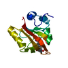
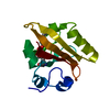
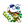
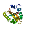
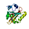
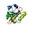
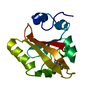
| Title | PYP Ultra-high resolution structure of a Bacterial Photoreceptor | ||||||
 Components Components | Photoactive yellow protein | ||||||
 Keywords Keywords | SIGNALING PROTEIN / PAS / LOV / GAF / domains fold | ||||||
| Function / homology |  Function and homology information Function and homology informationphotoreceptor activity / phototransduction / regulation of DNA-templated transcription / identical protein binding Similarity search - Function | ||||||
| Biological species |  Halorhodospira halophila (bacteria) Halorhodospira halophila (bacteria) | ||||||
| Method |  X-RAY DIFFRACTION / SYNCHROTRON / MOLECULAR REPLACEMENT / Resolution: 0.82 Å X-RAY DIFFRACTION / SYNCHROTRON / MOLECULAR REPLACEMENT / Resolution: 0.82 Å | ||||||
 Authors Authors | Getzoff, E.D. / Gutwin, K.N. / Genick, U.K. | ||||||
 Citation Citation | Journal: Nat.Struct.Biol. / Year: 2003 Title: Anticipatory active-site motions and chromophore distortions prime photoreceptor PYP for light activation Authors: Getzoff, E.D. / Gutwin, K.N. / Genick, U.K. | ||||||
| History |
|
- Structure visualization
| Structure viewer | Molecule: MolmilJmol/JSmol |
|---|
- Downloads & links
-Download
| PDBx/mmCIF format | 1nwz.cif.gz | 75.5 KB | Display | PDBx/mmCIF format |
|---|---|---|---|---|
| PDB format | pdb1nwz.ent.gz | 56.2 KB | Display | PDB format |
| PDBx/mmJSON format | 1nwz.json.gz | Tree view | PDBx/mmJSON format | |
| Others |  Other downloads Other downloads |
-Validation report
| Arichive directory | https://data.pdbj.org/pub/pdb/validation_reports/nw/1nwzftp://data.pdbj.org/pub/pdb/validation_reports/nw/1nwz | HTTPS FTP |
|---|
-Related structure data
| Similar structure data |
|---|


-Links
 PDBj
PDBj


- Assembly
| Deposited unit | 
| |||||||||||||||
|---|---|---|---|---|---|---|---|---|---|---|---|---|---|---|---|---|
| 1 |
| |||||||||||||||
| Unit cell |
| |||||||||||||||
| Components on special symmetry positions |
|
-Components
| #1: Protein | Mass: 13888.575 Da / Num. of mol.: 1 Source method: isolated from a genetically manipulated source Source: (gene. exp.) Halorhodospira halophila (bacteria) / Gene: PYP / Plasmid: pet20b / Species (production host): Escherichia coli / Production host: |
|---|---|
| #2: Chemical | ChemComp-HC4 /   Mass: 164.158 Da / Num. of mol.: 1 / Source method: obtained synthetically / Formula: C9H8O3 Mass: 164.158 Da / Num. of mol.: 1 / Source method: obtained synthetically / Formula: C9H8O3 |
| #3: Water | ChemComp-HOH /  Mass: 18.015 Da / Num. of mol.: 146 / Source method: isolated from a natural source / Formula: H2O Mass: 18.015 Da / Num. of mol.: 146 / Source method: isolated from a natural source / Formula: H2O |
| Has protein modification | Y |
-Experimental details
-Experiment
| Experiment | Method: X-RAY DIFFRACTION / Number of used crystals: 1 |
|---|
- Sample preparation
| Crystal | Density Matthews: 1.81 Å3/Da / Density % sol: 31.86 % |
|---|---|
| Crystal grow | Temperature: 298 K / Method: vapor diffusion, hanging drop / pH: 7 Details: ~2.5 M NH4SO4 20 mM Sodium Phosphate, pH 7, VAPOR DIFFUSION, HANGING DROP, temperature 298K |
| Crystal grow | *PLUS Method: unknown / Details: Genick, U.K., (1998) Nature, 392, 206. |
-Data collection
| Diffraction | Mean temperature: 149 K |
|---|---|
| Diffraction source | Source: SYNCHROTRON / Site: SSRL  / Beamline: BL9-1 / Wavelength: 0.77 Å / Beamline: BL9-1 / Wavelength: 0.77 Å |
| Detector | Type: MARRESEARCH / Detector: IMAGE PLATE / Date: Sep 6, 1997 |
| Radiation | Monochromator: Si(111) / Protocol: SINGLE WAVELENGTH / Monochromatic (M) / Laue (L): M / Scattering type: x-ray |
| Radiation wavelength | Wavelength: 0.77 Å / Relative weight: 1 |
| Reflection | Resolution: 0.82→30 Å / Num. all: 96611 / Num. obs: 96611 / % possible obs: 97.5 % / Observed criterion σ(F): 0 / Observed criterion σ(I): 0 |
| Reflection shell | Resolution: 0.82→0.83 Å / % possible all: 85.7 |
| Reflection | *PLUS Num. obs: 94197 / Redundancy: 4.5 % / Rmerge(I) obs: 0.031 |
| Reflection shell | *PLUS % possible obs: 85.7 % / Rmerge(I) obs: 0.442 / Mean I/σ(I) obs: 2.1 |
- Processing
| Software |
| |||||||||||||||||||||||||
|---|---|---|---|---|---|---|---|---|---|---|---|---|---|---|---|---|---|---|---|---|---|---|---|---|---|---|
| Refinement | Method to determine structure: MOLECULAR REPLACEMENT / Resolution: 0.82→30 Å / Cross valid method: THROUGHOUT / σ(F): 0 / σ(I): 0
| |||||||||||||||||||||||||
| Refinement step | Cycle: LAST / Resolution: 0.82→30 Å
| |||||||||||||||||||||||||
| Software | *PLUS Name: SHELXL / Version: 97 / Classification: refinement | |||||||||||||||||||||||||
| Refinement | *PLUS % reflection Rfree: 5 % / Rfactor Rfree: 0.1437 / Rfactor Rwork: 0.1232 | |||||||||||||||||||||||||
| Solvent computation | *PLUS | |||||||||||||||||||||||||
| Displacement parameters | *PLUS | |||||||||||||||||||||||||
| Refine LS restraints | *PLUS
|
