[English] 日本語
Yorodumi
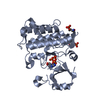
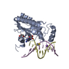
- PDB-6mgf: untagged, wild-type LptB in complex with ADP -

+
Open data


ID or keywords:

Loading...

-
Basic information

Entry
Database: PDB / ID: 6mgf
Titleuntagged, wild-type LptB in complex with ADP
ComponentsLipopolysaccharide export system ATP-binding protein LptB
KeywordsLIPID TRANSPORT / LptB / lipopolysaccharide / LPS transport / ABC-transporter / ATP-binding cassette / activator
Function / homology
Function and homology information


Translocases; Catalysing the translocation of carbohydrates and their derivatives; Linked to the hydrolysis of a nucleoside triphosphate / transporter complex / lipopolysaccharide transport / Gram-negative-bacterium-type cell outer membrane assembly / ATP-binding cassette (ABC) transporter complex / transmembrane transport / ATP hydrolysis activity / ATP binding / membrane / plasma membrane / cytoplasm
Similarity search - Function
Branched-chain amino acid ATP-binding cassette transporter, C-terminal / Branched-chain amino acid ATP-binding cassette transporter / LPS export ABC transporter, ATP-binding protein LptB / ABC transporter-like, conserved site / ABC transporters family signature. / ABC transporter / ABC transporter-like, ATP-binding domain / ATP-binding cassette, ABC transporter-type domain profile. / P-loop containing nucleotide triphosphate hydrolases / ATPases associated with a variety of cellular activities ...Branched-chain amino acid ATP-binding cassette transporter, C-terminal / Branched-chain amino acid ATP-binding cassette transporter / LPS export ABC transporter, ATP-binding protein LptB / ABC transporter-like, conserved site / ABC transporters family signature. / ABC transporter / ABC transporter-like, ATP-binding domain / ATP-binding cassette, ABC transporter-type domain profile. / P-loop containing nucleotide triphosphate hydrolases / ATPases associated with a variety of cellular activities / AAA+ ATPase domain / Rossmann fold / P-loop containing nucleoside triphosphate hydrolase / 3-Layer(aba) Sandwich / Alpha Beta
Similarity search - Domain/homology
ADENOSINE-5'-DIPHOSPHATE / Chem-CZJ / Lipopolysaccharide export system ATP-binding protein LptB
Similarity search - Component
Biological speciesEscherichia coli (E. coli)
MethodX-RAY DIFFRACTION / SYNCHROTRON / MOLECULAR REPLACEMENT / molecular replacement / Resolution: 2.982 Å
AuthorsMandler, M.D. / Owens, T.W. / Ruiz, N. / Kahne, D.
Funding support United States, 2items
OrganizationGrant numberCountry
National Institutes of Health/National Institute of General Medical Sciences (NIH/NIGMS)R01 GM066174 United States
National Institutes of Health/National Institute Of Allergy and Infectious Diseases (NIH/NIAID)R01 AI081059 United States
CitationJournal: not published
Title: Bypassing the requirement for coupling of ATP binding and hydrolysis in the lipopolysaccharide ABC transporte
Authors: Simpson, B. / Ruiz, N.
History
DepositionSep 13, 2018Deposition site: RCSB / Processing site: RCSB
Revision 1.0Sep 18, 2019Provider: repository / Type: Initial release
Revision 1.1Dec 18, 2019Group: Author supporting evidence / Category: pdbx_audit_support / Item: _pdbx_audit_support.funding_organization
Revision 1.2Oct 11, 2023Group: Data collection / Database references ...Data collection / Database references / Derived calculations / Refinement description / Structure summary
Category: chem_comp / chem_comp_atom ...chem_comp / chem_comp_atom / chem_comp_bond / citation / database_2 / entity / pdbx_entity_nonpoly / pdbx_initial_refinement_model
Item: _chem_comp.name / _citation.journal_abbrev ..._chem_comp.name / _citation.journal_abbrev / _database_2.pdbx_DOI / _database_2.pdbx_database_accession / _entity.pdbx_description / _pdbx_entity_nonpoly.name

-
Structure visualization

Structure viewerMolecule:
MolmilJmol/JSmol

Downloads & links

-
Assembly

Deposited unit
A: Lipopolysaccharide export system ATP-binding protein LptB
hetero molecules


Theoretical massNumber of molelcules
Total (without water)27,8764
Polymers26,8381
Non-polymers1,0383
Water1448
1


  • Idetical with deposited unit
  • defined by author&software
TypeNameSymmetry operationNumber
identity operation1_555x,y,z1
MethodPISA
Unit cell
Length a, b, c (Å)102.742, 34.767, 62.897
Angle α, β, γ (deg.)90.000, 100.070, 90.000
Int Tables number5
Space group name H-MC121

-
Components

#1: Protein Lipopolysaccharide export system ATP-binding protein LptB


Mass: 26837.668 Da / Num. of mol.: 1
Source method: isolated from a genetically manipulated source
Source: (gene. exp.) Escherichia coli (strain K12) (bacteria)
Strain: K12 / Gene: lptB, yhbG, b3201, JW3168 / Production host: Escherichia coli BL21(DE3) (bacteria)
References: UniProt: P0A9V1, Hydrolases; Acting on acid anhydrides; Acting on acid anhydrides to catalyse transmembrane movement of substances
#2: Chemical ChemComp-MG / MAGNESIUM ION


Mass: 24.305 Da / Num. of mol.: 1 / Source method: obtained synthetically / Formula: Mg
#3: Chemical ChemComp-ADP / ADENOSINE-5'-DIPHOSPHATE


Mass: 427.201 Da / Num. of mol.: 1 / Source method: obtained synthetically / Formula: C10H15N5O10P2 / Comment: ADP, energy-carrying molecule*YM
#4: Chemical ChemComp-CZJ / (3s,5s,7s)-N-{7-[(3-O-carbamoyl-6-deoxy-5-methyl-4-O-methyl-beta-D-gulopyranosyl)oxy]-4-hydroxy-8-methyl-2-oxo-2H-1-ben zopyran-3-yl}tricyclo[3.3.1.1~3,7~]decane-1-carboxamide / novobiocin derivative


Mass: 586.630 Da / Num. of mol.: 1 / Source method: obtained synthetically / Formula: C30H38N2O10
#5: Water ChemComp-HOH / water


Mass: 18.015 Da / Num. of mol.: 8 / Source method: isolated from a natural source / Formula: H2O

-
Experimental details

-
Experiment

ExperimentMethod: X-RAY DIFFRACTION / Number of used crystals: 1

-
Sample preparation

CrystalDensity Matthews: 2.07 Å3/Da / Density % sol: 40.6 %
Crystal growTemperature: 296 K / Method: liquid diffusion / pH: 6.5
Details: 22% w/v PEG 4000, 100mM MES pH 6.5, 1.3mM novobiocin-adamantyl, 7% DMSO

-
Data collection

DiffractionMean temperature: 100 K
Diffraction sourceSource: SYNCHROTRON / Site: APS / Beamline: 24-ID-E / Wavelength: 0.97918 Å
DetectorType: ADSC QUANTUM 315 / Detector: CCD / Date: Nov 17, 2016
RadiationProtocol: SINGLE WAVELENGTH / Monochromatic (M) / Laue (L): M / Scattering type: x-ray
Radiation wavelengthWavelength: 0.97918 Å / Relative weight: 1
ReflectionResolution: 2.98→61.93 Å / Num. obs: 4568 / % possible obs: 98.2 % / Redundancy: 5.2 % / Biso Wilson estimate: 40.25 Å2 / CC1/2: 0.967 / Rmerge(I) obs: 0.294 / Rpim(I) all: 0.137 / Rrim(I) all: 0.326 / Net I/σ(I): 6.4 / Num. measured all: 23772 / Scaling rejects: 4
Reflection shell

Diffraction-ID: 1

Resolution (Å)Redundancy (%)Rmerge(I) obsNum. unique obsCC1/2Rpim(I) allRrim(I) all% possible all
2.98-3.164.41.0936720.5910.5531.23189.4
8.95-61.934.80.0721900.9940.0360.08199.7

-
Phasing

PhasingMethod: molecular replacement
Phasing MR
Highest resolutionLowest resolution
Rotation5.31 Å61.93 Å
Translation5.31 Å61.93 Å

-
Processing

Software
NameVersionClassification
Aimless0.5.29data scaling
PHASER2.8.1phasing
PHENIX1.13_2998refinement
PDB_EXTRACT3.24data extraction
XDSdata reduction
RefinementMethod to determine structure: MOLECULAR REPLACEMENT
Starting model: 6B8B
Resolution: 2.982→61.928 Å / SU ML: 0.42 / Cross valid method: THROUGHOUT / σ(F): 1.34 / Phase error: 21.71
RfactorNum. reflection% reflection
Rfree0.2367 456 9.99 %
Rwork0.2012 --
obs0.2048 4566 98.05 %
Solvent computationShrinkage radii: 0.9 Å / VDW probe radii: 1.11 Å
Displacement parametersBiso max: 103.58 Å2 / Biso mean: 42.2853 Å2 / Biso min: 4.97 Å2
Refinement stepCycle: final / Resolution: 2.982→61.928 Å
ProteinNucleic acidLigandSolventTotal
Num. atoms1855 0 70 8 1933
Biso mean--61.48 17.12 -
Num. residues----240
LS refinement shell

Refine-ID: X-RAY DIFFRACTION / Rfactor Rfree error: 0 / Total num. of bins used: 3

Resolution (Å)Rfactor RfreeNum. reflection RfreeRfactor RworkNum. reflection RworkNum. reflection all% reflection obs (%)
2.982-3.41340.30271450.25921303144894
3.4134-4.30040.23631540.193313811535100
4.3004-61.94120.20431570.178714261583100
Refinement TLS params.

Method: refined / Refine-ID: X-RAY DIFFRACTION

IDL112)L122)L132)L222)L232)L332)S11 (Å °)S12 (Å °)S13 (Å °)S21 (Å °)S22 (Å °)S23 (Å °)S31 (Å °)S32 (Å °)S33 (Å °)T112)T122)T132)T222)T232)T332)Origin x (Å)Origin y (Å)Origin z (Å)
15.5757-0.6777-0.78894.36631.09984.53690.12170.26680.1388-0.0253-0.03710.5758-0.3809-0.4204-0.2620.2765-0.0476-0.04460.2470.03820.2289-22.1254-15.447711.445
21.5678-1.26990.8081.49830.15053.0977-0.15930.0628-0.28910.31790.01580.20330.0135-0.31460.10880.2229-0.06390.02360.27080.06760.2492-18.9044-14.402914.4117
34.62150.5079-0.48562.47540.80113.44720.2012-0.5666-0.94780.3167-0.4278-0.17970.2658-0.13440.19530.284-0.050.07410.30240.07230.3441-16.6001-25.775519.3733
43.3428-1.6131.99312.1522-0.92295.03210.4228-0.1938-0.3572-0.0593-0.24990.26450.23580.5276-0.18120.3752-0.1262-0.02060.6301-0.06860.41190.0546-20.676127.3085
55.7932-1.44771.55991.5531.32622.9070.48530.3027-0.4934-0.14090.4602-0.84981.22771.8296-0.52970.66810.3088-0.02470.8034-0.25770.88967.9259-27.8523.0562
61.4371-2.13252.68793.676-3.2626.2742-0.41540.44560.39660.21290.0911-0.6408-1.05820.76840.1530.4225-0.1778-0.02380.98680.05440.336110.0243-13.42524.5364
72.04710.9230.59181.8151-1.95974.09020.0650.0053-0.01630.157-0.2063-0.5303-0.19320.7080.17980.2494-0.01410.01120.3727-0.030.4098-0.223-14.822113.6394
83.0345-2.16112.5393.5634-0.43062.99460.1078-0.28720.30260.1023-0.1178-0.385-0.1689-0.0458-0.02930.2889-0.07080.0620.39630.01720.2419-9.4445-11.76124.4626
92.7516-1.4355-1.90811.90230.16433.385-0.08310.3802-0.3913-0.1311-0.21370.26250.0955-0.94840.1440.204-0.0041-0.07580.41320.07030.32-18.4555-9.7921-1.9796
109.85564.4837-8.79712.0429-4.00397.86021.3166-0.42491.22030.879-0.6763-0.2359-1.05860.572-0.0980.65250.1494-0.09750.49730.01830.4458-15.42071.2562-3.6945
Refinement TLS group
IDRefine-IDRefine TLS-IDSelection detailsAuth asym-IDAuth seq-ID
1X-RAY DIFFRACTION1chain 'A' and (resid 2 through 30 )A2 - 30
2X-RAY DIFFRACTION2chain 'A' and (resid 31 through 62 )A31 - 62
3X-RAY DIFFRACTION3chain 'A' and (resid 63 through 83 )A63 - 83
4X-RAY DIFFRACTION4chain 'A' and (resid 84 through 110 )A84 - 110
5X-RAY DIFFRACTION5chain 'A' and (resid 111 through 124 )A111 - 124
6X-RAY DIFFRACTION6chain 'A' and (resid 125 through 139 )A125 - 139
7X-RAY DIFFRACTION7chain 'A' and (resid 140 through 185 )A140 - 185
8X-RAY DIFFRACTION8chain 'A' and (resid 186 through 211 )A186 - 211
9X-RAY DIFFRACTION9chain 'A' and (resid 212 through 227 )A212 - 227
10X-RAY DIFFRACTION10chain 'A' and (resid 228 through 241 )A228 - 241

+
About Yorodumi

-
News

-
Feb 9, 2022. New format data for meta-information of EMDB entries

New format data for meta-information of EMDB entries

  • Version 3 of the EMDB header file is now the official format.
  • The previous official version 1.9 will be removed from the archive.

Related info.:EMDB header

External links:wwPDB to switch to version 3 of the EMDB data model

-
Aug 12, 2020. Covid-19 info

Covid-19 info

URL: https://pdbjlvh1.pdbj.org/emnavi/covid19.php

New page: Covid-19 featured information page in EM Navigator.

Related info.:Covid-19 info / Mar 5, 2020. Novel coronavirus structure data

+
Mar 5, 2020. Novel coronavirus structure data

Novel coronavirus structure data

Related info.:Yorodumi Speices / Aug 12, 2020. Covid-19 info

External links:COVID-19 featured content - PDBj / Molecule of the Month (242):Coronavirus Proteases

+
Jan 31, 2019. EMDB accession codes are about to change! (news from PDBe EMDB page)

EMDB accession codes are about to change! (news from PDBe EMDB page)

  • The allocation of 4 digits for EMDB accession codes will soon come to an end. Whilst these codes will remain in use, new EMDB accession codes will include an additional digit and will expand incrementally as the available range of codes is exhausted. The current 4-digit format prefixed with “EMD-” (i.e. EMD-XXXX) will advance to a 5-digit format (i.e. EMD-XXXXX), and so on. It is currently estimated that the 4-digit codes will be depleted around Spring 2019, at which point the 5-digit format will come into force.
  • The EM Navigator/Yorodumi systems omit the EMD- prefix.

Related info.:Q: What is EMD? / ID/Accession-code notation in Yorodumi/EM Navigator

External links:EMDB Accession Codes are Changing Soon! / Contact to PDBj

+
Jul 12, 2017. Major update of PDB

Major update of PDB

  • wwPDB released updated PDB data conforming to the new PDBx/mmCIF dictionary.
  • This is a major update changing the version number from 4 to 5, and with Remediation, in which all the entries are updated.
  • In this update, many items about electron microscopy experimental information are reorganized (e.g. em_software).
  • Now, EM Navigator and Yorodumi are based on the updated data.

External links:wwPDB Remediation / Enriched Model Files Conforming to OneDep Data Standards Now Available in the PDB FTP Archive

-
Yorodumi

Thousand views of thousand structures

  • Yorodumi is a browser for structure data from EMDB, PDB, SASBDB, etc.
  • This page is also the successor to EM Navigator detail page, and also detail information page/front-end page for Omokage search.
  • The word "yorodu" (or yorozu) is an old Japanese word meaning "ten thousand". "mi" (miru) is to see.

Related info.:EMDB / PDB / SASBDB / Comparison of 3 databanks / Yorodumi Search / Aug 31, 2016. New EM Navigator & Yorodumi / Yorodumi Papers / Jmol/JSmol / Function and homology information / Changes in new EM Navigator and Yorodumi

Read more