 Movie
Movie Controller

[English] 日本語
 Yorodumi
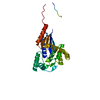
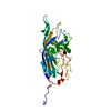
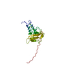
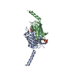
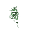
- PDB-6mf5: Crystal structure of budding yeast Cdc5 polo-box domain in comple... -
+ Open data

- Basic information
| Entry | Database: PDB / ID: 6mf5 | ||||||
|---|---|---|---|---|---|---|---|
| Title | Crystal structure of budding yeast Cdc5 polo-box domain in complex with Spc72 phosphopeptide. | ||||||
 Components Components |
| ||||||
 Keywords Keywords | CELL CYCLE / Transferase / Mitosis / Dbf4 / Polo-like kinases | ||||||
| Function / homology |  Function and homology information Function and homology informationgamma-tubulin complex localization to cytoplasmic side of mitotic spindle pole body / astral microtubule anchoring at mitotic spindle pole body / outer plaque of mitotic spindle pole body / positive regulation of mitotic spindle pole body separation / positive regulation of protein localization to cell division site involved in mitotic actomyosin contractile ring assembly / regulation of protein localization to mitotic spindle pole body / regulation of nucleus organization / Polo-like kinase mediated events / Regulation of PLK1 Activity at G2/M Transition / TP53 regulates transcription of additional cell cycle genes whose exact role in the p53 pathway remain uncertain ...gamma-tubulin complex localization to cytoplasmic side of mitotic spindle pole body / astral microtubule anchoring at mitotic spindle pole body / outer plaque of mitotic spindle pole body / positive regulation of mitotic spindle pole body separation / positive regulation of protein localization to cell division site involved in mitotic actomyosin contractile ring assembly / regulation of protein localization to mitotic spindle pole body / regulation of nucleus organization / Polo-like kinase mediated events / Regulation of PLK1 Activity at G2/M Transition / TP53 regulates transcription of additional cell cycle genes whose exact role in the p53 pathway remain uncertain / mitotic spindle orientation checkpoint signaling / Golgi Cisternae Pericentriolar Stack Reorganization / outer plaque of spindle pole body / Resolution of Sister Chromatid Cohesion / negative regulation of protein localization to nucleolus / meiotic spindle assembly / regulation of protein localization to nucleolus / mitotic spindle astral microtubule end / mitotic spindle pole body / synaptonemal complex disassembly / karyogamy / polo kinase / centromeric DNA binding / resolution of meiotic recombination intermediates / astral microtubule organization / cellular bud neck / microtubule nucleation / microtubule plus-end binding / spindle pole body / exit from mitosis / microtubule lateral binding / Ubiquitin-Mediated Degradation of Phosphorylated Cdc25A / positive regulation of Rho protein signal transduction / cytoplasmic microtubule organization / mitotic spindle organization / spindle microtubule / phosphoprotein binding / kinetochore / G2/M transition of mitotic cell cycle / spindle pole / protein kinase activity / protein serine kinase activity / cell division / protein serine/threonine kinase activity / protein-containing complex binding / ATP binding / nucleus / cytoplasm Similarity search - Function | ||||||
| Biological species |  | ||||||
| Method |  X-RAY DIFFRACTION / SYNCHROTRON / MOLECULAR REPLACEMENT / Resolution: 2.7 Å X-RAY DIFFRACTION / SYNCHROTRON / MOLECULAR REPLACEMENT / Resolution: 2.7 Å | ||||||
 Authors Authors | Guarne, A. / Almawi, A.W. | ||||||
| Funding support |  Canada, 1items Canada, 1items
| ||||||
 Citation Citation | Journal: Sci Rep / Year: 2020 Title: Distinct surfaces on Cdc5/PLK Polo-box domain orchestrate combinatorial substrate recognition during cell division. Authors: Almawi, A.W. / Langlois-Lemay, L. / Boulton, S. / Rodriguez Gonzalez, J. / Melacini, G. / D'Amours, D. / Guarne, A. | ||||||
| History |
|
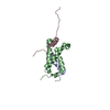
- Structure visualization
| Structure viewer | Molecule: MolmilJmol/JSmol |
|---|
- Downloads & links
-Download
| PDBx/mmCIF format | 6mf5.cif.gz | 282.9 KB | Display | PDBx/mmCIF format |
|---|---|---|---|---|
| PDB format | pdb6mf5.ent.gz | 231.4 KB | Display | PDB format |
| PDBx/mmJSON format | 6mf5.json.gz | Tree view | PDBx/mmJSON format | |
| Others |  Other downloads Other downloads |
-Validation report
| Arichive directory | https://data.pdbj.org/pub/pdb/validation_reports/mf/6mf5ftp://data.pdbj.org/pub/pdb/validation_reports/mf/6mf5 | HTTPS FTP |
|---|
-Related structure data
| Related structure data |  6mf4C  6mf6C  1q4oS S: Starting model for refinement C: citing same article ( |
|---|---|
| Similar structure data |

-Links
 PDBj
PDBj


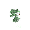
- Assembly
| Deposited unit | 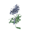
| ||||||||
|---|---|---|---|---|---|---|---|---|---|
| 1 | 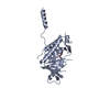
| ||||||||
| 2 | 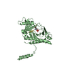
| ||||||||
| Unit cell |
|
-Components
| #1: Protein | Mass: 33463.184 Da / Num. of mol.: 2 Source method: isolated from a genetically manipulated source Source: (gene. exp.) Strain: ATCC 204508 / S288c / Gene: CDC5, MSD2, PKX2, YMR001C, YM8270.03C / Production host:  #2: Protein/peptide | Mass: 1112.043 Da / Num. of mol.: 2 Source method: isolated from a genetically manipulated source Source: (gene. exp.) Production host: synthetic construct (others) / References: UniProt: P39723*PLUS #3: Chemical |   Mass: 35.453 Da / Num. of mol.: 2 / Source method: obtained synthetically / Formula: Cl Mass: 35.453 Da / Num. of mol.: 2 / Source method: obtained synthetically / Formula: Cl#4: Water | ChemComp-HOH / |  Mass: 18.015 Da / Num. of mol.: 11 / Source method: isolated from a natural source / Formula: H2O Mass: 18.015 Da / Num. of mol.: 11 / Source method: isolated from a natural source / Formula: H2OHas protein modification | Y | |
|---|
-Experimental details
-Experiment
| Experiment | Method: X-RAY DIFFRACTION / Number of used crystals: 1 |
|---|
- Sample preparation
| Crystal | Density Matthews: 2.14 Å3/Da / Density % sol: 42.54 % |
|---|---|
| Crystal grow | Temperature: 277 K / Method: vapor diffusion, sitting drop Details: 100 mM Bis-Tris pH 6.50, 200 mM magnesium chloride, 100 mM cesium chloride, 25 % PEG 3350. |
-Data collection
| Diffraction | Mean temperature: 100 K |
|---|---|
| Diffraction source | Source: SYNCHROTRON / Site: APS  / Beamline: 17-ID / Wavelength: 0.9795 Å / Beamline: 17-ID / Wavelength: 0.9795 Å |
| Detector | Type: DECTRIS PILATUS3 S 6M / Detector: PIXEL / Date: Dec 13, 2016 |
| Radiation | Protocol: SINGLE WAVELENGTH / Monochromatic (M) / Laue (L): M / Scattering type: x-ray |
| Radiation wavelength | Wavelength: 0.9795 Å / Relative weight: 1 |
| Reflection | Resolution: 2.7→42.1 Å / Num. obs: 16158 / % possible obs: 99.9 % / Redundancy: 5.8 % / CC1/2: 0.99 / Net I/σ(I): 9.1 |
| Reflection shell | Resolution: 2.7→2.78 Å / Num. unique obs: 1184 / CC1/2: 0.317 |
- Processing
| Software |
| |||||||||||||||||||||||||||||||||||||||||||||||||||||||||||||||||||||||||||||||||||||||||||||||||||||||||||||||||||||||||||||||||||||||||||||||||||||||||||||||||||||||||||||||
|---|---|---|---|---|---|---|---|---|---|---|---|---|---|---|---|---|---|---|---|---|---|---|---|---|---|---|---|---|---|---|---|---|---|---|---|---|---|---|---|---|---|---|---|---|---|---|---|---|---|---|---|---|---|---|---|---|---|---|---|---|---|---|---|---|---|---|---|---|---|---|---|---|---|---|---|---|---|---|---|---|---|---|---|---|---|---|---|---|---|---|---|---|---|---|---|---|---|---|---|---|---|---|---|---|---|---|---|---|---|---|---|---|---|---|---|---|---|---|---|---|---|---|---|---|---|---|---|---|---|---|---|---|---|---|---|---|---|---|---|---|---|---|---|---|---|---|---|---|---|---|---|---|---|---|---|---|---|---|---|---|---|---|---|---|---|---|---|---|---|---|---|---|---|---|---|---|
| Refinement | Method to determine structure: MOLECULAR REPLACEMENT Starting model: 1Q4O Resolution: 2.7→42.08 Å / SU ML: 0.34 / Cross valid method: FREE R-VALUE / σ(F): 1.34 / Phase error: 30.73
| |||||||||||||||||||||||||||||||||||||||||||||||||||||||||||||||||||||||||||||||||||||||||||||||||||||||||||||||||||||||||||||||||||||||||||||||||||||||||||||||||||||||||||||||
| Solvent computation | Shrinkage radii: 0.9 Å / VDW probe radii: 1.11 Å | |||||||||||||||||||||||||||||||||||||||||||||||||||||||||||||||||||||||||||||||||||||||||||||||||||||||||||||||||||||||||||||||||||||||||||||||||||||||||||||||||||||||||||||||
| Refinement step | Cycle: LAST / Resolution: 2.7→42.08 Å
| |||||||||||||||||||||||||||||||||||||||||||||||||||||||||||||||||||||||||||||||||||||||||||||||||||||||||||||||||||||||||||||||||||||||||||||||||||||||||||||||||||||||||||||||
| Refine LS restraints |
| |||||||||||||||||||||||||||||||||||||||||||||||||||||||||||||||||||||||||||||||||||||||||||||||||||||||||||||||||||||||||||||||||||||||||||||||||||||||||||||||||||||||||||||||
| LS refinement shell |
| |||||||||||||||||||||||||||||||||||||||||||||||||||||||||||||||||||||||||||||||||||||||||||||||||||||||||||||||||||||||||||||||||||||||||||||||||||||||||||||||||||||||||||||||
| Refinement TLS params. | Method: refined / Refine-ID: X-RAY DIFFRACTION
| |||||||||||||||||||||||||||||||||||||||||||||||||||||||||||||||||||||||||||||||||||||||||||||||||||||||||||||||||||||||||||||||||||||||||||||||||||||||||||||||||||||||||||||||
| Refinement TLS group |
|
