 Movie
Movie Controller


+ Open data

- Basic information
| Entry | Database: PDB / ID: 6m7y | |||||||||
|---|---|---|---|---|---|---|---|---|---|---|
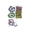
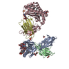
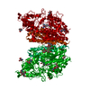
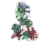
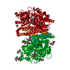
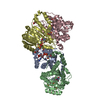
| Title | Dehydratase, NisB, bound to a non-eliminable substrate analog | |||||||||
 Components Components |
| |||||||||
 Keywords Keywords | BIOSYNTHETIC PROTEIN / RiPP / nisin / tRNA-dependent / dehydratase | |||||||||
| Function / homology |  Function and homology information Function and homology informationkilling of cells of another organism / defense response to bacterium / signaling receptor binding / extracellular region / plasma membrane Similarity search - Function | |||||||||
| Biological species |  Lactococcus lactis subsp. lactis (lactic acid bacteria)Lactococcus lactis (lactic acid bacteria) Lactococcus lactis subsp. lactis (lactic acid bacteria)Lactococcus lactis (lactic acid bacteria) | |||||||||
| Method |  X-RAY DIFFRACTION / SYNCHROTRON / MOLECULAR REPLACEMENT / Resolution: 2.794 Å X-RAY DIFFRACTION / SYNCHROTRON / MOLECULAR REPLACEMENT / Resolution: 2.794 Å | |||||||||
 Authors Authors | Cogan, D.P. / Nair, S.K. | |||||||||
 Citation Citation | Journal: Proc.Natl.Acad.Sci.USA / Year: 2019 Title: Characterization of glutamyl-tRNA-dependent dehydratases using nonreactive substrate mimics. Authors: Bothwell, I.R. / Cogan, D.P. / Kim, T. / Reinhardt, C.J. / van der Donk, W.A. / Nair, S.K. | |||||||||
| History |
|
- Structure visualization
| Structure viewer | Molecule: MolmilJmol/JSmol |
|---|
- Downloads & links
-Download
| PDBx/mmCIF format | 6m7y.cif.gz | 419.3 KB | Display | PDBx/mmCIF format |
|---|---|---|---|---|
| PDB format | pdb6m7y.ent.gz | 336.6 KB | Display | PDB format |
| PDBx/mmJSON format | 6m7y.json.gz | Tree view | PDBx/mmJSON format | |
| Others |  Other downloads Other downloads |
-Validation report
| Arichive directory | https://data.pdbj.org/pub/pdb/validation_reports/m7/6m7yftp://data.pdbj.org/pub/pdb/validation_reports/m7/6m7y | HTTPS FTP |
|---|
-Related structure data
| Related structure data | 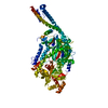 6ec7C 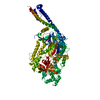 6ec8C 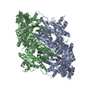 4wd9S S: Starting model for refinement C: citing same article ( |
|---|---|
| Similar structure data |
-Links
 PDBj
PDBj- Assembly
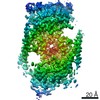
| Deposited unit | 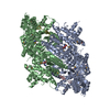
| ||||||||
|---|---|---|---|---|---|---|---|---|---|
| 1 | 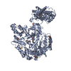
| ||||||||
| 2 | 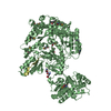
| ||||||||
| Unit cell |
|
-Components
| #1: Protein | Mass: 118647.109 Da / Num. of mol.: 2 / Mutation: V169C Source method: isolated from a genetically manipulated source Source: (gene. exp.) Lactococcus lactis subsp. lactis (lactic acid bacteria)Gene: nisB / Plasmid: pET28b-NisB-V169C / Production host: #2: Protein/peptide | Mass: 3641.135 Da / Num. of mol.: 2 / Mutation: S12C, S26(J9A), C30A, C34A / Source method: obtained synthetically / Source: (synth.) Lactococcus lactis (lactic acid bacteria) / References: UniProt: Q7BB86, UniProt: P29559*PLUS#3: Water | ChemComp-HOH / |  Mass: 18.015 Da / Num. of mol.: 247 / Source method: isolated from a natural source / Formula: H2O Mass: 18.015 Da / Num. of mol.: 247 / Source method: isolated from a natural source / Formula: H2OHas protein modification | Y | |
|---|
-Experimental details
-Experiment
| Experiment | Method: X-RAY DIFFRACTION / Number of used crystals: 1 |
|---|
- Sample preparation
| Crystal | Density Matthews: 2.8 Å3/Da / Density % sol: 56.14 % |
|---|---|
| Crystal grow | Temperature: 282 K / Method: vapor diffusion, hanging drop / pH: 8 Details: 18.3% PEG 6000, 0.1 M bicine pH 8.0, 1 mM dithiothreitol |
-Data collection
| Diffraction | Mean temperature: 100 K / Serial crystal experiment: N |
|---|---|
| Diffraction source | Source: SYNCHROTRON / Site: APS  / Beamline: 21-ID-G / Wavelength: 0.97856 Å / Beamline: 21-ID-G / Wavelength: 0.97856 Å |
| Detector | Type: MARMOSAIC 300 mm CCD / Detector: CCD / Date: Jun 2, 2017 |
| Radiation | Protocol: SINGLE WAVELENGTH / Monochromatic (M) / Laue (L): M / Scattering type: x-ray |
| Radiation wavelength | Wavelength: 0.97856 Å / Relative weight: 1 |
| Reflection | Resolution: 2.79→127.61 Å / Num. obs: 62069 / % possible obs: 93.1 % / Redundancy: 6.9 % / CC1/2: 0.998 / Rmerge(I) obs: 0.09 / Net I/σ(I): 21.5 |
| Reflection shell | Resolution: 2.79→2.89 Å / Rmerge(I) obs: 0.836 / Num. unique obs: 4205 / CC1/2: 0.867 |
- Processing
| Software |
| |||||||||||||||||||||||||||||||||||||||||||||||||||||||||||||||||||||||||||||||||||||||||||||||||||||||||||||||||||||||||||||||||||||||||||||||||||||||||||||||||
|---|---|---|---|---|---|---|---|---|---|---|---|---|---|---|---|---|---|---|---|---|---|---|---|---|---|---|---|---|---|---|---|---|---|---|---|---|---|---|---|---|---|---|---|---|---|---|---|---|---|---|---|---|---|---|---|---|---|---|---|---|---|---|---|---|---|---|---|---|---|---|---|---|---|---|---|---|---|---|---|---|---|---|---|---|---|---|---|---|---|---|---|---|---|---|---|---|---|---|---|---|---|---|---|---|---|---|---|---|---|---|---|---|---|---|---|---|---|---|---|---|---|---|---|---|---|---|---|---|---|---|---|---|---|---|---|---|---|---|---|---|---|---|---|---|---|---|---|---|---|---|---|---|---|---|---|---|---|---|---|---|---|---|
| Refinement | Method to determine structure: MOLECULAR REPLACEMENT Starting model: 4WD9 Resolution: 2.794→29.393 Å / SU ML: 0.4 / Cross valid method: THROUGHOUT / σ(F): 1.35 / Phase error: 28.79
| |||||||||||||||||||||||||||||||||||||||||||||||||||||||||||||||||||||||||||||||||||||||||||||||||||||||||||||||||||||||||||||||||||||||||||||||||||||||||||||||||
| Solvent computation | Shrinkage radii: 0.9 Å / VDW probe radii: 1.11 Å | |||||||||||||||||||||||||||||||||||||||||||||||||||||||||||||||||||||||||||||||||||||||||||||||||||||||||||||||||||||||||||||||||||||||||||||||||||||||||||||||||
| Refinement step | Cycle: LAST / Resolution: 2.794→29.393 Å
| |||||||||||||||||||||||||||||||||||||||||||||||||||||||||||||||||||||||||||||||||||||||||||||||||||||||||||||||||||||||||||||||||||||||||||||||||||||||||||||||||
| Refine LS restraints |
| |||||||||||||||||||||||||||||||||||||||||||||||||||||||||||||||||||||||||||||||||||||||||||||||||||||||||||||||||||||||||||||||||||||||||||||||||||||||||||||||||
| LS refinement shell |
|
