 Movie
Movie Controller

[English] 日本語
 Yorodumi
- PDB-3a22: Crystal Structure of beta-L-Arabinopyranosidase complexed with L-... -
+ Open data

- Basic information
| Entry | Database: PDB / ID: 3a22 | ||||||
|---|---|---|---|---|---|---|---|
| Title | Crystal Structure of beta-L-Arabinopyranosidase complexed with L-arabinose | ||||||
 Components Components | Putative secreted alpha-galactosidase | ||||||
 Keywords Keywords | HYDROLASE / beta-alpha-barrel / Greek key motif / beta-jellyroll / beta-trefoil | ||||||
| Function / homology |  Function and homology information Function and homology informationalpha-galactosidase / alpha-galactosidase activity / carbohydrate metabolic process Similarity search - Function | ||||||
| Biological species |  Streptomyces avermitilis (bacteria) Streptomyces avermitilis (bacteria) | ||||||
| Method |  X-RAY DIFFRACTION / SYNCHROTRON / MOLECULAR REPLACEMENT / Resolution: 1.9 Å X-RAY DIFFRACTION / SYNCHROTRON / MOLECULAR REPLACEMENT / Resolution: 1.9 Å | ||||||
 Authors Authors | Fujimoto, Z. / Ichinose, H. / Kaneko, S. | ||||||
 Citation Citation | Journal: J.Biol.Chem. / Year: 2009 Title: A beta-l-Arabinopyranosidase from Streptomyces avermitilis is a novel member of glycoside hydrolase family 27. Authors: Ichinose, H. / Fujimoto, Z. / Honda, M. / Harazono, K. / Nishimoto, Y. / Uzura, A. / Kaneko, S. #1: Journal: Acta Crystallogr.,Sect.F / Year: 2008 Title: Crystallization and preliminary crystallographic analysis of exo-alpha-1,5-L-arabinofuranosidase from Streptomyces avermitilis NBRC14893. Authors: Fujimoto, Z. / Ichinose, H. / Kaneko, S. | ||||||
| History |
|
- Structure visualization
| Structure viewer | Molecule: MolmilJmol/JSmol |
|---|
- Downloads & links
-Download
| PDBx/mmCIF format | 3a22.cif.gz | 276.1 KB | Display | PDBx/mmCIF format |
|---|---|---|---|---|
| PDB format | pdb3a22.ent.gz | 218.2 KB | Display | PDB format |
| PDBx/mmJSON format | 3a22.json.gz | Tree view | PDBx/mmJSON format | |
| Others |  Other downloads Other downloads |
-Validation report
| Arichive directory | https://data.pdbj.org/pub/pdb/validation_reports/a2/3a22ftp://data.pdbj.org/pub/pdb/validation_reports/a2/3a22 | HTTPS FTP |
|---|
-Related structure data
| Related structure data |  3a21SC  3a23C S: Starting model for refinement C: citing same article ( |
|---|---|
| Similar structure data |


-Links
 PDBj
PDBj

- Assembly
| Deposited unit | 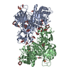
| ||||||||
|---|---|---|---|---|---|---|---|---|---|
| 1 | 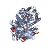
| ||||||||
| 2 | 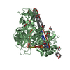
| ||||||||
| Unit cell |
|
-Components
-Protein / Sugars , 2 types, 11 molecules AB
| #1: Protein | Mass: 64307.629 Da / Num. of mol.: 2 / Fragment: UNP residues 45-658 Source method: isolated from a genetically manipulated source Source: (gene. exp.) Streptomyces avermitilis (bacteria) / Strain: NBRC14893 / Gene: agaA2, SAV2186, SAV_2186 / Plasmid: pIJ702 / Production host: Streptomyces lividans (bacteria) / Strain (production host): 1326 / References: UniProt: Q82L26#2: Sugar | ChemComp-ARA /  Type: L-saccharide, alpha linking / Mass: 150.130 Da / Num. of mol.: 9 Type: L-saccharide, alpha linking / Mass: 150.130 Da / Num. of mol.: 9Source method: isolated from a genetically manipulated source Formula: C5H10O5 |
|---|
-Non-polymers , 5 types, 1386 molecules 


| #3: Chemical | ChemComp-GOL /  Mass: 92.094 Da / Num. of mol.: 18 / Source method: obtained synthetically / Formula: C3H8O3 Mass: 92.094 Da / Num. of mol.: 18 / Source method: obtained synthetically / Formula: C3H8O3#4: Chemical | ChemComp-1PG /  Mass: 252.305 Da / Num. of mol.: 4 / Source method: obtained synthetically / Formula: C11H24O6 Mass: 252.305 Da / Num. of mol.: 4 / Source method: obtained synthetically / Formula: C11H24O6#5: Chemical |  Mass: 96.063 Da / Num. of mol.: 3 / Source method: obtained synthetically / Formula: SO4 Mass: 96.063 Da / Num. of mol.: 3 / Source method: obtained synthetically / Formula: SO4#6: Chemical | ChemComp-EPE / |  Mass: 238.305 Da / Num. of mol.: 1 / Source method: obtained synthetically / Formula: C8H18N2O4S / Comment: pH buffer*YM Mass: 238.305 Da / Num. of mol.: 1 / Source method: obtained synthetically / Formula: C8H18N2O4S / Comment: pH buffer*YM#7: Water | ChemComp-HOH / | Mass: 18.015 Da / Num. of mol.: 1360 / Source method: isolated from a natural source / Formula: H2O |
|---|
-Details
| Has protein modification | Y |
|---|
-Experimental details
-Experiment
| Experiment | Method: X-RAY DIFFRACTION / Number of used crystals: 1 |
|---|
- Sample preparation
| Crystal | Density Matthews: 2.4 Å3/Da / Density % sol: 48.76 % |
|---|---|
| Crystal grow | Temperature: 293 K / Method: vapor diffusion, sitting drop / pH: 8 Details: 1.7M ammonium sulfate, 1.7% PEG 400, 15% glycerol, 0.1M HEPES, pH 8.0, VAPOR DIFFUSION, SITTING DROP, temperature 293K |
-Data collection
| Diffraction | Mean temperature: 95 K |
|---|---|
| Diffraction source | Source: SYNCHROTRON / Site: Photon Factory  / Beamline: BL-6A / Wavelength: 0.978 Å / Beamline: BL-6A / Wavelength: 0.978 Å |
| Detector | Type: ADSC QUANTUM 4 / Detector: CCD / Date: Nov 25, 2008 |
| Radiation | Protocol: SINGLE WAVELENGTH / Monochromatic (M) / Laue (L): M / Scattering type: x-ray |
| Radiation wavelength | Wavelength: 0.978 Å / Relative weight: 1 |
| Reflection | Resolution: 1.9→50 Å / Num. obs: 97305 / % possible obs: 99.9 % / Observed criterion σ(I): 0 / Redundancy: 9 % / Rmerge(I) obs: 0.075 / Net I/σ(I): 31.7 |
| Reflection shell | Resolution: 1.9→1.97 Å / Redundancy: 8.7 % / Rmerge(I) obs: 0.23 / Mean I/σ(I) obs: 12.2 / % possible all: 100 |
- Processing
| Software |
| ||||||||||||||||||||||||||||||||||||||||||||||||||||||||||||||||||||||||||||||||||||||||||||||||||||||||||||||||||||||||||||||||||||||||||||||||||||||||||||||||||||||||||
|---|---|---|---|---|---|---|---|---|---|---|---|---|---|---|---|---|---|---|---|---|---|---|---|---|---|---|---|---|---|---|---|---|---|---|---|---|---|---|---|---|---|---|---|---|---|---|---|---|---|---|---|---|---|---|---|---|---|---|---|---|---|---|---|---|---|---|---|---|---|---|---|---|---|---|---|---|---|---|---|---|---|---|---|---|---|---|---|---|---|---|---|---|---|---|---|---|---|---|---|---|---|---|---|---|---|---|---|---|---|---|---|---|---|---|---|---|---|---|---|---|---|---|---|---|---|---|---|---|---|---|---|---|---|---|---|---|---|---|---|---|---|---|---|---|---|---|---|---|---|---|---|---|---|---|---|---|---|---|---|---|---|---|---|---|---|---|---|---|---|---|---|
| Refinement | Method to determine structure: MOLECULAR REPLACEMENT Starting model: PDB ENTRY 3A21 Resolution: 1.9→41.27 Å / Cor.coef. Fo:Fc: 0.953 / Cor.coef. Fo:Fc free: 0.923 / SU B: 3.015 / SU ML: 0.088 / Cross valid method: THROUGHOUT / ESU R: 0.149 / ESU R Free: 0.139 / Stereochemistry target values: MAXIMUM LIKELIHOOD / Details: HYDROGENS HAVE BEEN ADDED IN THE RIDING POSITIONS
| ||||||||||||||||||||||||||||||||||||||||||||||||||||||||||||||||||||||||||||||||||||||||||||||||||||||||||||||||||||||||||||||||||||||||||||||||||||||||||||||||||||||||||
| Solvent computation | Ion probe radii: 0.8 Å / Shrinkage radii: 0.8 Å / VDW probe radii: 1.2 Å / Solvent model: MASK | ||||||||||||||||||||||||||||||||||||||||||||||||||||||||||||||||||||||||||||||||||||||||||||||||||||||||||||||||||||||||||||||||||||||||||||||||||||||||||||||||||||||||||
| Displacement parameters | Biso mean: 13.101 Å2
| ||||||||||||||||||||||||||||||||||||||||||||||||||||||||||||||||||||||||||||||||||||||||||||||||||||||||||||||||||||||||||||||||||||||||||||||||||||||||||||||||||||||||||
| Refine analyze |
| ||||||||||||||||||||||||||||||||||||||||||||||||||||||||||||||||||||||||||||||||||||||||||||||||||||||||||||||||||||||||||||||||||||||||||||||||||||||||||||||||||||||||||
| Refinement step | Cycle: LAST / Resolution: 1.9→41.27 Å
| ||||||||||||||||||||||||||||||||||||||||||||||||||||||||||||||||||||||||||||||||||||||||||||||||||||||||||||||||||||||||||||||||||||||||||||||||||||||||||||||||||||||||||
| Refine LS restraints |
| ||||||||||||||||||||||||||||||||||||||||||||||||||||||||||||||||||||||||||||||||||||||||||||||||||||||||||||||||||||||||||||||||||||||||||||||||||||||||||||||||||||||||||
| LS refinement shell | Resolution: 1.9→1.949 Å / Total num. of bins used: 20
|
