 Movie
Movie Controller

[English] 日本語
 Yorodumi
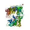
- PDB-6m31: Structural and Functional Insights into an Archaeal Lipid Synthase -
+ Open data

- Basic information
| Entry | Database: PDB / ID: 6m31 | ||||||
|---|---|---|---|---|---|---|---|
| Title | Structural and Functional Insights into an Archaeal Lipid Synthase | ||||||
 Components Components | Digeranylgeranylglyceryl phosphate synthase | ||||||
 Keywords Keywords | MEMBRANE PROTEIN / Lipid | ||||||
| Function / homology |  Function and homology information Function and homology informationgeranylgeranylglycerol-phosphate geranylgeranyltransferase / geranylgeranylglycerol-phosphate geranylgeranyltransferase activity / glycerophospholipid biosynthetic process / magnesium ion binding / plasma membrane Similarity search - Function | ||||||
| Biological species |   Methanocaldococcus jannaschii DSM 2661 (archaea) Methanocaldococcus jannaschii DSM 2661 (archaea) | ||||||
| Method |  X-RAY DIFFRACTION / SYNCHROTRON / SAD / Resolution: 2.3 Å X-RAY DIFFRACTION / SYNCHROTRON / SAD / Resolution: 2.3 Å | ||||||
 Authors Authors | Ren, S. / Cheng, W. | ||||||
 Citation Citation | Journal: Cell Rep / Year: 2020 Title: Structural and Functional Insights into an Archaeal Lipid Synthase Authors: Ren, S. / de Kok, N.A. / Gu, Y. / Yan, W. / Sun, Q. / Chen, Y. / He, J. / Tian, L. / Andringa, R.L. / Zhu, X. / Tang, M. / Qi, S. / Xu, H. / Ren, H. / Fu, X. / Minnaard, A.J. / Yang, S. / ...Authors: Ren, S. / de Kok, N.A. / Gu, Y. / Yan, W. / Sun, Q. / Chen, Y. / He, J. / Tian, L. / Andringa, R.L. / Zhu, X. / Tang, M. / Qi, S. / Xu, H. / Ren, H. / Fu, X. / Minnaard, A.J. / Yang, S. / Zhang, W. / Li, W. / Wei, Y. / Driessen, A.J. / Cheng, W. | ||||||
| History |
|
- Structure visualization
| Structure viewer | Molecule: MolmilJmol/JSmol |
|---|
- Downloads & links
-Download
| PDBx/mmCIF format | 6m31.cif.gz | 143.3 KB | Display | PDBx/mmCIF format |
|---|---|---|---|---|
| PDB format | pdb6m31.ent.gz | 115.9 KB | Display | PDB format |
| PDBx/mmJSON format | 6m31.json.gz | Tree view | PDBx/mmJSON format | |
| Others |  Other downloads Other downloads |
-Validation report
| Arichive directory | https://data.pdbj.org/pub/pdb/validation_reports/m3/6m31ftp://data.pdbj.org/pub/pdb/validation_reports/m3/6m31 | HTTPS FTP |
|---|
-Related structure data


-Links
 PDBj
PDBj
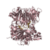
- Assembly
| Deposited unit | 
| ||||||||
|---|---|---|---|---|---|---|---|---|---|
| 1 |
| ||||||||
| Unit cell |
|
-Components
-Protein , 1 types, 2 molecules AB
| #1: Protein | Mass: 31785.689 Da / Num. of mol.: 2 Source method: isolated from a genetically manipulated source Source: (gene. exp.) Methanocaldococcus jannaschii DSM 2661 (archaea)Strain: DSM 2661 / Gene: MJ0279 / Production host:  References: UniProt: Q57727, geranylgeranylglycerol-phosphate geranylgeranyltransferase |
|---|
-Non-polymers , 5 types, 91 molecules 


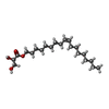

| #2: Chemical | ChemComp-LDA /  Mass: 229.402 Da / Num. of mol.: 23 / Source method: obtained synthetically / Formula: C14H31NO / Feature type: SUBJECT OF INVESTIGATION / Comment: LDAO, detergent*YM Mass: 229.402 Da / Num. of mol.: 23 / Source method: obtained synthetically / Formula: C14H31NO / Feature type: SUBJECT OF INVESTIGATION / Comment: LDAO, detergent*YM#3: Chemical | ChemComp-PO4 /  Mass: 94.971 Da / Num. of mol.: 5 / Source method: obtained synthetically / Formula: PO4 / Feature type: SUBJECT OF INVESTIGATION Mass: 94.971 Da / Num. of mol.: 5 / Source method: obtained synthetically / Formula: PO4 / Feature type: SUBJECT OF INVESTIGATION#4: Chemical |  Mass: 24.305 Da / Num. of mol.: 2 / Source method: obtained synthetically / Formula: Mg / Feature type: SUBJECT OF INVESTIGATION Mass: 24.305 Da / Num. of mol.: 2 / Source method: obtained synthetically / Formula: Mg / Feature type: SUBJECT OF INVESTIGATION#5: Chemical | ChemComp-MPG / [(  Mass: 356.540 Da / Num. of mol.: 18 / Source method: obtained synthetically / Formula: C21H40O4 / Feature type: SUBJECT OF INVESTIGATION Mass: 356.540 Da / Num. of mol.: 18 / Source method: obtained synthetically / Formula: C21H40O4 / Feature type: SUBJECT OF INVESTIGATION#6: Water | ChemComp-HOH / | Mass: 18.015 Da / Num. of mol.: 43 / Source method: isolated from a natural source / Formula: H2O |
|---|
-Details
| Has ligand of interest | Y |
|---|
-Experimental details
-Experiment
| Experiment | Method: X-RAY DIFFRACTION / Number of used crystals: 1 |
|---|
- Sample preparation
| Crystal | Density Matthews: 2.92 Å3/Da / Density % sol: 57.9 % |
|---|---|
| Crystal grow | Temperature: 293.15 K / Method: lipidic cubic phase Details: 200 mM NaCl, 20% (w/v) PEG500, and 200 mM Tris-HCl (pH 8.0). |
-Data collection
| Diffraction | Mean temperature: 100 K / Serial crystal experiment: N | ||||||||||||||||||||||||||||||||||||||||||||||||||||||||
|---|---|---|---|---|---|---|---|---|---|---|---|---|---|---|---|---|---|---|---|---|---|---|---|---|---|---|---|---|---|---|---|---|---|---|---|---|---|---|---|---|---|---|---|---|---|---|---|---|---|---|---|---|---|---|---|---|---|
| Diffraction source | Source: SYNCHROTRON / Site: SSRF  / Beamline: BL19U1 / Wavelength: 0.9793 Å / Beamline: BL19U1 / Wavelength: 0.9793 Å | ||||||||||||||||||||||||||||||||||||||||||||||||||||||||
| Detector | Type: DECTRIS PILATUS3 R 1M / Detector: PIXEL / Date: May 10, 2018 | ||||||||||||||||||||||||||||||||||||||||||||||||||||||||
| Radiation | Protocol: SINGLE WAVELENGTH / Monochromatic (M) / Laue (L): M / Scattering type: x-ray | ||||||||||||||||||||||||||||||||||||||||||||||||||||||||
| Radiation wavelength | Wavelength: 0.9793 Å / Relative weight: 1 | ||||||||||||||||||||||||||||||||||||||||||||||||||||||||
| Reflection | Resolution: 2.29→48.89 Å / Num. obs: 33780 / % possible obs: 99.8 % / Redundancy: 6.904 % / Biso Wilson estimate: 45.73 Å2 / CC1/2: 0.999 / Rmerge(I) obs: 0.092 / Rrim(I) all: 0.099 / Χ2: 1.221 / Net I/σ(I): 13.4 | ||||||||||||||||||||||||||||||||||||||||||||||||||||||||
| Reflection shell | Diffraction-ID: 1
|
- Processing
| Software |
| ||||||||||||||||||||||||
|---|---|---|---|---|---|---|---|---|---|---|---|---|---|---|---|---|---|---|---|---|---|---|---|---|---|
| Refinement | Method to determine structure: SAD / Resolution: 2.3→48.89 Å / Cor.coef. Fo:Fc: 0.942 / Cor.coef. Fo:Fc free: 0.939 / SU R Cruickshank DPI: 0.302 / Cross valid method: THROUGHOUT / σ(F): 0 / SU R Blow DPI: 0.289 / SU Rfree Blow DPI: 0.204 / SU Rfree Cruickshank DPI: 0.21
| ||||||||||||||||||||||||
| Displacement parameters | Biso max: 145.39 Å2 / Biso mean: 54.73 Å2 / Biso min: 20 Å2
| ||||||||||||||||||||||||
| Refine analyze | Luzzati coordinate error obs: 0.3 Å | ||||||||||||||||||||||||
| Refinement step | Cycle: final / Resolution: 2.3→48.89 Å
| ||||||||||||||||||||||||
| LS refinement shell | Resolution: 2.3→2.32 Å / Rfactor Rfree error: 0 / Total num. of bins used: 50
|
