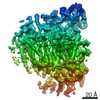
 Movie
Movie Controller

[English] 日本語
 Yorodumi
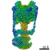
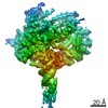
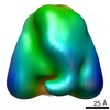
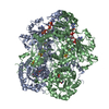
- PDB-6ly9: The membrane-embedded Vo domain of V/A-ATPase from Thermus thermo... -
+ Open data

- Basic information
| Entry | Database: PDB / ID: 6ly9 | ||||||||||||
|---|---|---|---|---|---|---|---|---|---|---|---|---|---|
| Title | The membrane-embedded Vo domain of V/A-ATPase from Thermus thermophilus | ||||||||||||
 Components Components |
| ||||||||||||
 Keywords Keywords | MOTOR PROTEIN / rotary ATPase / V/A-ATPase / molecular motor | ||||||||||||
| Function / homology |  Function and homology information Function and homology informationproton-transporting two-sector ATPase complex, catalytic domain / proton-transporting V-type ATPase, V0 domain / vacuolar proton-transporting V-type ATPase complex / vacuolar acidification / proton motive force-driven plasma membrane ATP synthesis / proton-transporting ATPase activity, rotational mechanism / proton-transporting ATP synthase activity, rotational mechanism / ATPase binding / ATP binding / metal ion binding Similarity search - Function | ||||||||||||
| Biological species |   Thermus thermophilus HB8 (bacteria) Thermus thermophilus HB8 (bacteria) | ||||||||||||
| Method | ELECTRON MICROSCOPY / single particle reconstruction / cryo EM / Resolution: 3.93 Å | ||||||||||||
 Authors Authors | Kishikawa, J. / Nakanishi, A. / Furuta, A. / Kato, T. / Namba, K. / Tamakoshi, M. / Mitsuoka, K. / Yokoyama, K. | ||||||||||||
| Funding support |  Japan, 3items Japan, 3items
| ||||||||||||
 Citation Citation | Journal: Elife / Year: 2020 Title: Mechanical inhibition of isolated V from V/A-ATPase for proton conductance. Authors: Jun-Ichi Kishikawa / Atsuko Nakanishi / Aya Furuta / Takayuki Kato / Keiichi Namba / Masatada Tamakoshi / Kaoru Mitsuoka / Ken Yokoyama / Abstract: V-ATPase is an energy converting enzyme, coupling ATP hydrolysis/synthesis in the hydrophilic V domain, with proton flow through the V membrane domain, via rotation of the central rotor complex ...V-ATPase is an energy converting enzyme, coupling ATP hydrolysis/synthesis in the hydrophilic V domain, with proton flow through the V membrane domain, via rotation of the central rotor complex relative to the surrounding stator apparatus. Upon dissociation from the V domain, the V domain of the eukaryotic V-ATPase can adopt a physiologically relevant auto-inhibited form in which proton conductance through the V domain is prevented, however the molecular mechanism of this inhibition is not fully understood. Using cryo-electron microscopy, we determined the structure of both the V/A-ATPase and isolated V at near-atomic resolution, respectively. These structures clarify how the isolated V domain adopts the auto-inhibited form and how the complex prevents formation of the inhibited V form. | ||||||||||||
| History |
|
- Structure visualization
| Movie |
Movie viewer |
|---|---|
| Structure viewer | Molecule: MolmilJmol/JSmol |
- Downloads & links
-Download
| PDBx/mmCIF format | 6ly9.cif.gz | 319.3 KB | Display | PDBx/mmCIF format |
|---|---|---|---|---|
| PDB format | pdb6ly9.ent.gz | 253.4 KB | Display | PDB format |
| PDBx/mmJSON format | 6ly9.json.gz | Tree view | PDBx/mmJSON format | |
| Others |  Other downloads Other downloads |
-Validation report
| Arichive directory | https://data.pdbj.org/pub/pdb/validation_reports/ly/6ly9ftp://data.pdbj.org/pub/pdb/validation_reports/ly/6ly9 | HTTPS FTP |
|---|
-Related structure data
| Related structure data |  30015MC  6ly8C M: map data used to model this data C: citing same article ( |
|---|---|
| Similar structure data |


-Links
 PDBj
PDBj

- Assembly
| Deposited unit | 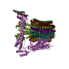
|
|---|---|
| 1 |
|
-Components
| #1: Protein | Mass: 72204.289 Da / Num. of mol.: 1 / Source method: isolated from a natural source / Source: (natural) Thermus thermophilus HB8 (bacteria) / Strain: HB8 / References: UniProt: Q5SIT6 | ||||||||
|---|---|---|---|---|---|---|---|---|---|
| #2: Protein | Mass: 9841.714 Da / Num. of mol.: 12 / Source method: isolated from a natural source / Source: (natural) Thermus thermophilus HB8 (bacteria) / Strain: HB8 / References: UniProt: Q5SIT7#3: Protein | | Mass: 35968.570 Da / Num. of mol.: 1 / Source method: isolated from a natural source / Source: (natural) Thermus thermophilus HB8 (bacteria) / Strain: HB8 / References: UniProt: P74902#4: Protein | | Mass: 13166.218 Da / Num. of mol.: 1 / Source method: isolated from a natural source / Source: (natural) Thermus thermophilus HB8 (bacteria) / Strain: HB8 / References: UniProt: Q5SIT5#5: Protein | | Mass: 20645.582 Da / Num. of mol.: 1 / Source method: isolated from a natural source / Source: (natural) Thermus thermophilus HB8 (bacteria) / Strain: HB8 / References: UniProt: P74901Has protein modification | Y | |
-Experimental details
-Experiment
| Experiment | Method: ELECTRON MICROSCOPY |
|---|---|
| EM experiment | Aggregation state: PARTICLE / 3D reconstruction method: single particle reconstruction |
- Sample preparation
| Component | Name: Membrane-embedded Vo domain / Type: COMPLEX / Details: V/A-type ATPase from Thermus thermophilus / Entity ID: all / Source: NATURAL | |||||||||||||||
|---|---|---|---|---|---|---|---|---|---|---|---|---|---|---|---|---|
| Molecular weight |
| |||||||||||||||
| Source (natural) | Organism: Thermus thermophilus HB8 (bacteria) | |||||||||||||||
| Buffer solution | pH: 8 | |||||||||||||||
| Buffer component |
| |||||||||||||||
| Specimen | Conc.: 3 mg/ml / Embedding applied: NO / Shadowing applied: NO / Staining applied: NO / Vitrification applied: YES Details: The sample was purified from cell membrane of Thermus thermophilus and incorporated into nanodisc. | |||||||||||||||
| Specimen support | Grid material: MOLYBDENUM / Grid mesh size: 300 divisions/in. / Grid type: Quantifoil R1.2/1.3 | |||||||||||||||
| Vitrification | Instrument: FEI VITROBOT MARK IV / Cryogen name: ETHANE / Humidity: 100 % / Chamber temperature: 277 K |
- Electron microscopy imaging
| Microscopy | Model: JEOL CRYO ARM 200 |
|---|---|
| Electron gun | Electron source:  FIELD EMISSION GUN / Accelerating voltage: 200 kV / Illumination mode: FLOOD BEAM FIELD EMISSION GUN / Accelerating voltage: 200 kV / Illumination mode: FLOOD BEAM |
| Electron lens | Mode: BRIGHT FIELD / Nominal magnification: 50000 X / Calibrated defocus min: 1000 nm / Calibrated defocus max: 2000 nm / Cs: 1.4 mm |
| Specimen holder | Cryogen: NITROGEN |
| Image recording | Average exposure time: 12 sec. / Electron dose: 79.2 e/Å2 / Detector mode: COUNTING / Film or detector model: GATAN K2 SUMMIT (4k x 4k) / Num. of real images: 5988 |
| EM imaging optics | Energyfilter name: In-column Omega Filter |
| Image scans | Width: 4096 / Height: 4096 / Movie frames/image: 60 / Used frames/image: 1-60 |
| EM diffraction | Camera length: 800 mm |
- Processing
| Software | Name: PHENIX / Classification: refinement | ||||||||||||||||||||||||||||||||
|---|---|---|---|---|---|---|---|---|---|---|---|---|---|---|---|---|---|---|---|---|---|---|---|---|---|---|---|---|---|---|---|---|---|
| EM software |
| ||||||||||||||||||||||||||||||||
| CTF correction | Type: PHASE FLIPPING AND AMPLITUDE CORRECTION | ||||||||||||||||||||||||||||||||
| Particle selection | Num. of particles selected: 3140000 Details: The particles selected from manually-selected 3268 micrographs. | ||||||||||||||||||||||||||||||||
| Symmetry | Point symmetry: C1 (asymmetric) | ||||||||||||||||||||||||||||||||
| 3D reconstruction | Resolution: 3.93 Å / Resolution method: FSC 0.143 CUT-OFF / Num. of particles: 157618 / Algorithm: FOURIER SPACE / Symmetry type: POINT | ||||||||||||||||||||||||||||||||
| Atomic model building | Protocol: AB INITIO MODEL / Space: REAL / Target criteria: Correlation coefficient | ||||||||||||||||||||||||||||||||
| Atomic model building | PDB-ID: 6QUM Accession code: 6QUM / Source name: PDB / Type: experimental model | ||||||||||||||||||||||||||||||||
| Refine LS restraints |
|

