Movie
Movie Controller

[English] 日本語
 Yorodumi
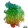
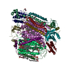
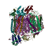
- EMDB-4720: Neurospora crassa cytochrome C oxidase (complex IV) in lipid nanodisc -
+ Open data

- Basic information
| Entry | Database: EMDB / ID: EMD-4720 | |||||||||
|---|---|---|---|---|---|---|---|---|---|---|
| Title | Neurospora crassa cytochrome C oxidase (complex IV) in lipid nanodisc | |||||||||
 Map data Map data | None | |||||||||
 Sample Sample |
| |||||||||
| Biological species |  Neurospora crassa (fungus) Neurospora crassa (fungus) | |||||||||
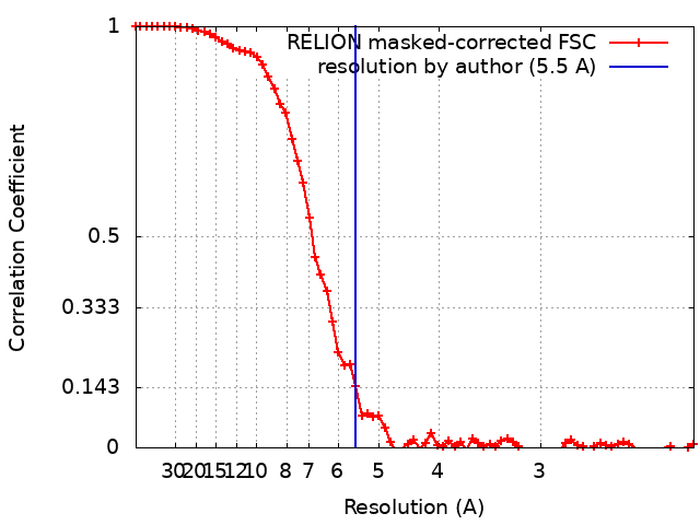
| Method | single particle reconstruction / cryo EM / Resolution: 5.5 Å | |||||||||
 Authors Authors | Bausewein T / Nussberger S / Kuehlbrandt W | |||||||||
| Funding support |  Germany, 1 items Germany, 1 items
| |||||||||
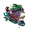
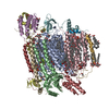
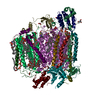
 Citation Citation | Journal: IUCrJ / Year: 2019 Title: Cryo-EM structure of respiratory complex IV. Authors: Thomas Bausewein / Stephan Nussberger / Werner Kühlbrandt / Abstract: In fungi, the mitochondrial respiratory chain complexes (complexes I-IV) are responsible for oxidative phosphorylation, as in higher eukaryotes. Cryo-EM was used to identify a 200 kDa membrane ...In fungi, the mitochondrial respiratory chain complexes (complexes I-IV) are responsible for oxidative phosphorylation, as in higher eukaryotes. Cryo-EM was used to identify a 200 kDa membrane protein from in lipid nanodiscs as cytochrome oxidase (complex IV) and its structure was determined at 5.5 Å resolution. The map closely resembles the cryo-EM structure of complex IV from . Its ten subunits are conserved in and , but other transmembrane subunits are missing. The different structure of the Cox5a subunit is typical for fungal complex IV and may affect the interaction with complex III in a respiratory supercomplex. Additional density was found between the matrix domains of the Cox4 and Cox5a subunits that appears to be specific to . | |||||||||
| History |
|
- Structure visualization
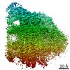
| Movie |
 Movie viewer Movie viewer |
|---|---|
| Structure viewer | EM map: SurfViewMolmilJmol/JSmol |
| Supplemental images |

- Downloads & links
-EMDB archive
| Map data | emd_4720.map.gz | 24.9 MB | EMDB map data format | |
|---|---|---|---|---|
| Header (meta data) | emd-4720-v30.xmlemd-4720.xml | 9.4 KB 9.4 KB | Display Display | EMDB header |
| FSC (resolution estimation) | emd_4720_fsc.xml | 7 KB | Display | FSC data file |
| Images | 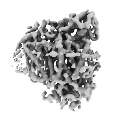 emd_4720.png emd_4720.png | 56.4 KB | ||
| Archive directory |  http://ftp.pdbj.org/pub/emdb/structures/EMD-4720ftp://ftp.pdbj.org/pub/emdb/structures/EMD-4720 http://ftp.pdbj.org/pub/emdb/structures/EMD-4720ftp://ftp.pdbj.org/pub/emdb/structures/EMD-4720 | HTTPS FTP |
-Related structure data
| Related structure data | |
|---|---|
| Similar structure data |


-Links
| EMDB pages | EMDB (EBI/PDBe) / EMDataResource |
|---|
-Map
| File | Download / File: emd_4720.map.gz / Format: CCP4 / Size: 27 MB / Type: IMAGE STORED AS FLOATING POINT NUMBER (4 BYTES) | ||||||||||||||||||||||||||||||||||||||||||||||||||||||||||||
|---|---|---|---|---|---|---|---|---|---|---|---|---|---|---|---|---|---|---|---|---|---|---|---|---|---|---|---|---|---|---|---|---|---|---|---|---|---|---|---|---|---|---|---|---|---|---|---|---|---|---|---|---|---|---|---|---|---|---|---|---|---|
| Annotation | None | ||||||||||||||||||||||||||||||||||||||||||||||||||||||||||||
| Projections & slices | Image control
Images are generated by Spider. | ||||||||||||||||||||||||||||||||||||||||||||||||||||||||||||
| Voxel size | X=Y=Z: 1.09 Å | ||||||||||||||||||||||||||||||||||||||||||||||||||||||||||||
| Density |
| ||||||||||||||||||||||||||||||||||||||||||||||||||||||||||||
| Symmetry | Space group: 1 | ||||||||||||||||||||||||||||||||||||||||||||||||||||||||||||
| Details | EMDB XML:
CCP4 map header:
| ||||||||||||||||||||||||||||||||||||||||||||||||||||||||||||
 Z (Sec.)
 Y (Row.)
 X (Col.)


-Supplemental data
- Sample components
-Entire : Complex IV, cytochrome c oxidase
| Entire | Name: Complex IV, cytochrome c oxidase |
|---|---|
| Components |
|
-Supramolecule #1: Complex IV, cytochrome c oxidase
| Supramolecule | Name: Complex IV, cytochrome c oxidase / type: complex / ID: 1 / Parent: 0 |
|---|---|
| Source (natural) | Organism: Neurospora crassa (fungus) / Organelle: Mitochondrium |
-Experimental details
-Structure determination
| Method | cryo EM |
|---|---|
 Processing Processing | single particle reconstruction |
| Aggregation state | particle |
-Sample preparation
| Buffer | pH: 7.2 |
|---|---|
| Vitrification | Cryogen name: ETHANE / Chamber humidity: 100 % / Chamber temperature: 283 K / Instrument: FEI VITROBOT MARK III |
- Electron microscopy
| Microscope | FEI POLARA 300 |
|---|---|
| Specialist optics | Energy filter - Slit width: 70 eV |
| Image recording | Film or detector model: GATAN K2 SUMMIT (4k x 4k) / Detector mode: COUNTING / Average electron dose: 2.0 e/Å2 |
| Electron beam | Acceleration voltage: 300 kV / Electron source:  FIELD EMISSION GUN FIELD EMISSION GUN |
| Electron optics | Illumination mode: FLOOD BEAM / Imaging mode: BRIGHT FIELD / Cs: 2.0 mm |
| Experimental equipment |  Model: Tecnai Polara / Image courtesy: FEI Company |