 Movie
Movie Controller

[English] 日本語
 Yorodumi
- PDB-6ltf: Dimeric isocitrate dehydrogenase from Xanthomonas campestris pv. ... -
+ Open data

- Basic information
| Entry | Database: PDB / ID: 6ltf | ||||||
|---|---|---|---|---|---|---|---|
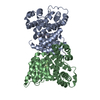
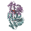
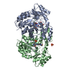
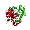
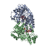
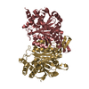
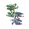
| Title | Dimeric isocitrate dehydrogenase from Xanthomonas campestris pv. campestris 8004 | ||||||
 Components Components | Isocitrate dehydrogenase | ||||||
 Keywords Keywords | OXIDOREDUCTASE / isocitrate dehydrogenase / BIOSYNTHETIC PROTEIN | ||||||
| Function / homology |  Function and homology information Function and homology informationisocitrate dehydrogenase (NAD+) / isocitrate dehydrogenase (NAD+) activity / oxidoreductase activity, acting on the CH-OH group of donors, NAD or NADP as acceptor / NAD binding / magnesium ion binding Similarity search - Function | ||||||
| Biological species |  Xanthomonas campestris (bacteria) Xanthomonas campestris (bacteria) | ||||||
| Method |  X-RAY DIFFRACTION / SYNCHROTRON / MOLECULAR REPLACEMENT / Resolution: 1.61 Å X-RAY DIFFRACTION / SYNCHROTRON / MOLECULAR REPLACEMENT / Resolution: 1.61 Å | ||||||
 Authors Authors | Zhu, G.P. | ||||||
 Citation Citation | Journal: To Be Published Title: Dimeric isocitrate dehydrogenase from Xanthomonas campestris pv. campestris 8004 Authors: Zhu, G.P. | ||||||
| History |
|
- Structure visualization
| Structure viewer | Molecule: MolmilJmol/JSmol |
|---|
- Downloads & links
-Download
| PDBx/mmCIF format | 6ltf.cif.gz | 103.5 KB | Display | PDBx/mmCIF format |
|---|---|---|---|---|
| PDB format | pdb6ltf.ent.gz | 63.6 KB | Display | PDB format |
| PDBx/mmJSON format | 6ltf.json.gz | Tree view | PDBx/mmJSON format | |
| Others |  Other downloads Other downloads |
-Validation report
| Arichive directory | https://data.pdbj.org/pub/pdb/validation_reports/lt/6ltfftp://data.pdbj.org/pub/pdb/validation_reports/lt/6ltf | HTTPS FTP |
|---|
-Related structure data
| Related structure data |  6m3sC 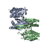 5grhS S: Starting model for refinement C: citing same article ( |
|---|---|
| Similar structure data |


-Links
 PDBj
PDBj

- Assembly
| Deposited unit | 
| ||||||||||||
|---|---|---|---|---|---|---|---|---|---|---|---|---|---|
| 1 | 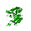
| ||||||||||||
| Unit cell |
|
-Components
| #1: Protein | Mass: 35516.504 Da / Num. of mol.: 1 Source method: isolated from a genetically manipulated source Source: (gene. exp.) Xanthomonas campestris (bacteria) / Gene: D0A35_19175, TR80_00940, XcmpCFBP7700_13825 / Production host: References: UniProt: A0A2S4JWD6, UniProt: A0A0H2XBX7*PLUS, isocitrate dehydrogenase (NAD+) |
|---|---|
| #2: Chemical | ChemComp-BEZ /   Mass: 122.121 Da / Num. of mol.: 1 / Source method: obtained synthetically / Formula: C7H6O2 Mass: 122.121 Da / Num. of mol.: 1 / Source method: obtained synthetically / Formula: C7H6O2 |
| #3: Water | ChemComp-HOH /  Mass: 18.015 Da / Num. of mol.: 387 / Source method: isolated from a natural source / Formula: H2O Mass: 18.015 Da / Num. of mol.: 387 / Source method: isolated from a natural source / Formula: H2O |
| Has ligand of interest | N |
-Experimental details
-Experiment
| Experiment | Method: X-RAY DIFFRACTION / Number of used crystals: 1 |
|---|
- Sample preparation
| Crystal | Density Matthews: 2.27 Å3/Da / Density % sol: 45.85 % |
|---|---|
| Crystal grow | Temperature: 295 K / Method: vapor diffusion, hanging drop / pH: 8.5 / Details: 0.1 M Tris-HCl pH 8.5,0.2 M LiSO4, 10% Glycerol |
-Data collection
| Diffraction | Mean temperature: 100 K / Serial crystal experiment: N |
|---|---|
| Diffraction source | Source: SYNCHROTRON / Site: APS  / Beamline: 8-BM / Wavelength: 0.9791 Å / Beamline: 8-BM / Wavelength: 0.9791 Å |
| Detector | Type: DECTRIS PILATUS 6M-F / Detector: PIXEL / Date: May 24, 2016 |
| Radiation | Protocol: SINGLE WAVELENGTH / Monochromatic (M) / Laue (L): M / Scattering type: x-ray |
| Radiation wavelength | Wavelength: 0.9791 Å / Relative weight: 1 |
| Reflection | Resolution: 1.6→50 Å / Num. obs: 40051 / % possible obs: 96 % / Redundancy: 5.4 % / Biso Wilson estimate: 16.08 Å2 / Rmerge(I) obs: 0.103 / Rpim(I) all: 0.047 / Net I/σ(I): 24.5 |
| Reflection shell | Resolution: 1.6→1.63 Å / Redundancy: 5.2 % / Rmerge(I) obs: 0.542 / Mean I/σ(I) obs: 3 / Num. unique obs: 1993 / CC1/2: 0.814 / Rpim(I) all: 0.255 / % possible all: 98.3 |
- Processing
| Software |
| |||||||||||||||||||||||||||||||||||||||||||||||||||||||||||||||||||||||||||||||||||||||||||||||||||||||||
|---|---|---|---|---|---|---|---|---|---|---|---|---|---|---|---|---|---|---|---|---|---|---|---|---|---|---|---|---|---|---|---|---|---|---|---|---|---|---|---|---|---|---|---|---|---|---|---|---|---|---|---|---|---|---|---|---|---|---|---|---|---|---|---|---|---|---|---|---|---|---|---|---|---|---|---|---|---|---|---|---|---|---|---|---|---|---|---|---|---|---|---|---|---|---|---|---|---|---|---|---|---|---|---|---|---|---|
| Refinement | Method to determine structure: MOLECULAR REPLACEMENT Starting model: 5GRH Resolution: 1.61→43.42 Å / SU ML: 0.1583 / Cross valid method: FREE R-VALUE / σ(F): 0.15 / Phase error: 21.0084
| |||||||||||||||||||||||||||||||||||||||||||||||||||||||||||||||||||||||||||||||||||||||||||||||||||||||||
| Solvent computation | Shrinkage radii: 0.9 Å / VDW probe radii: 1.11 Å | |||||||||||||||||||||||||||||||||||||||||||||||||||||||||||||||||||||||||||||||||||||||||||||||||||||||||
| Displacement parameters | Biso mean: 22.78 Å2 | |||||||||||||||||||||||||||||||||||||||||||||||||||||||||||||||||||||||||||||||||||||||||||||||||||||||||
| Refinement step | Cycle: LAST / Resolution: 1.61→43.42 Å
| |||||||||||||||||||||||||||||||||||||||||||||||||||||||||||||||||||||||||||||||||||||||||||||||||||||||||
| Refine LS restraints |
| |||||||||||||||||||||||||||||||||||||||||||||||||||||||||||||||||||||||||||||||||||||||||||||||||||||||||
| LS refinement shell |
|
