 Movie
Movie Controller


+ Open data

- Basic information
| Entry | Database: PDB / ID: 6loj | ||||||
|---|---|---|---|---|---|---|---|
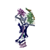
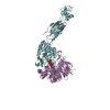
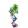
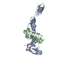
| Title | The complex structure of IpaH9.8-LRR and hGBP1 | ||||||
 Components Components |
| ||||||
 Keywords Keywords | LIGASE/HYDROLASE / IpaH9.8 / hGBP1 / LRR / TRANSFERASE / LIGASE-HYDROLASE complex | ||||||
| Function / homology |  Function and homology information Function and homology informationmodulation of process of another organism / GDP phosphatase activity / non-canonical inflammasome complex assembly / negative regulation of substrate adhesion-dependent cell spreading / protein localization to vacuole / symbiont cell surface / protein K27-linked ubiquitination / cytolysis in another organism / positive regulation of pyroptotic inflammatory response / vesicle membrane ...modulation of process of another organism / GDP phosphatase activity / non-canonical inflammasome complex assembly / negative regulation of substrate adhesion-dependent cell spreading / protein localization to vacuole / symbiont cell surface / protein K27-linked ubiquitination / cytolysis in another organism / positive regulation of pyroptotic inflammatory response / vesicle membrane / host cell cytosol / negative regulation of interleukin-2 production / negative regulation of T cell receptor signaling pathway / spectrin binding / defense response to protozoan / ligase activity / cytokine binding / Hydrolases; Acting on acid anhydrides; In phosphorus-containing anhydrides / cellular response to interleukin-1 / negative regulation of protein localization to plasma membrane / protein K48-linked ubiquitination / regulation of protein localization to plasma membrane / regulation of calcium-mediated signaling / cytoplasmic vesicle membrane / lipopolysaccharide binding / Hsp90 protein binding / negative regulation of ERK1 and ERK2 cascade / RING-type E3 ubiquitin transferase / cellular response to type II interferon / Interferon gamma signaling / ubiquitin-protein transferase activity / GDP binding / ubiquitin protein ligase activity / cellular response to tumor necrosis factor / actin cytoskeleton / actin binding / G protein activity / cytoplasmic vesicle / Hydrolases; Acting on acid anhydrides; Acting on GTP to facilitate cellular and subcellular movement / defense response to virus / host cell cytoplasm / protein ubiquitination / defense response to bacterium / symbiont-mediated suppression of host innate immune response / Golgi membrane / innate immune response / GTPase activity / GTP binding / host cell nucleus / enzyme binding / Golgi apparatus / protein homodimerization activity / extracellular region / identical protein binding / plasma membrane / cytosol / cytoplasm Similarity search - Function | ||||||
| Biological species |  Shigella flexneri (bacteria) Shigella flexneri (bacteria) Homo sapiens (human) Homo sapiens (human) | ||||||
| Method |  X-RAY DIFFRACTION / SYNCHROTRON / MOLECULAR REPLACEMENT / Resolution: 3.72 Å X-RAY DIFFRACTION / SYNCHROTRON / MOLECULAR REPLACEMENT / Resolution: 3.72 Å | ||||||
 Authors Authors | Ye, Y. / Huang, H. | ||||||
| Funding support |  China, 1items China, 1items
| ||||||
 Citation Citation | Journal: Commun Biol / Year: 2020 Title: Substrate-binding destabilizes the hydrophobic cluster to relieve the autoinhibition of bacterial ubiquitin ligase IpaH9.8. Authors: Ye, Y. / Xiong, Y. / Huang, H. | ||||||
| History |
|
- Structure visualization
| Structure viewer | Molecule: MolmilJmol/JSmol |
|---|
- Downloads & links
-Download
| PDBx/mmCIF format | 6loj.cif.gz | 335.8 KB | Display | PDBx/mmCIF format |
|---|---|---|---|---|
| PDB format | pdb6loj.ent.gz | 269.9 KB | Display | PDB format |
| PDBx/mmJSON format | 6loj.json.gz | Tree view | PDBx/mmJSON format | |
| Others |  Other downloads Other downloads |
-Validation report
| Arichive directory | https://data.pdbj.org/pub/pdb/validation_reports/lo/6lojftp://data.pdbj.org/pub/pdb/validation_reports/lo/6loj | HTTPS FTP |
|---|
-Related structure data
| Related structure data | 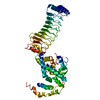 6lolC  1f5nS S: Starting model for refinement C: citing same article ( |
|---|---|
| Similar structure data |

-Links
 PDBj
PDBj- Assembly
| Deposited unit | 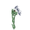
| ||||||||
|---|---|---|---|---|---|---|---|---|---|
| 1 |
| ||||||||
| Unit cell |
|
-Components
| #1: Protein | Mass: 25620.730 Da / Num. of mol.: 1 / Fragment: LRR domain Source method: isolated from a genetically manipulated source Source: (gene. exp.) Shigella flexneri (bacteria) / Gene: ipaH9.8, NCTC9783_05321 / Production host: References: UniProt: A0A380D7J9, UniProt: D2AJU0*PLUS, Ligases; Forming carbon-nitrogen bonds; Acid-amino-acid ligases (peptide synthases) |
|---|---|
| #2: Protein | Mass: 68375.047 Da / Num. of mol.: 1 / Mutation: Q507H Source method: isolated from a genetically manipulated source Source: (gene. exp.) Homo sapiens (human) / Gene: GBP1 / Production host: References: UniProt: P32455, Hydrolases; Acting on acid anhydrides; Acting on GTP to facilitate cellular and subcellular movement |
| #3: Chemical | ChemComp-GDP /   Type: RNA linking / Mass: 443.201 Da / Num. of mol.: 1 / Source method: obtained synthetically / Formula: C10H15N5O11P2 / Comment: GDP, energy-carrying molecule*YM Type: RNA linking / Mass: 443.201 Da / Num. of mol.: 1 / Source method: obtained synthetically / Formula: C10H15N5O11P2 / Comment: GDP, energy-carrying molecule*YM |
| Has ligand of interest | N |
-Experimental details
-Experiment
| Experiment | Method: X-RAY DIFFRACTION / Number of used crystals: 1 |
|---|
- Sample preparation
| Crystal | Density Matthews: 6.39 Å3/Da / Density % sol: 80.75 % |
|---|---|
| Crystal grow | Temperature: 298 K / Method: vapor diffusion, sitting drop / pH: 6 / Details: 2.5M NaCl, 0.1M K/Na phosphate buffer, pH 6 |
-Data collection
| Diffraction | Mean temperature: 100 K / Serial crystal experiment: N |
|---|---|
| Diffraction source | Source: SYNCHROTRON / Site: SSRF / Beamline: BL17U1 / Wavelength: 0.97 Å |
| Detector | Type: DECTRIS EIGER X 16M / Detector: PIXEL / Date: May 9, 2019 |
| Radiation | Protocol: SINGLE WAVELENGTH / Monochromatic (M) / Laue (L): M / Scattering type: x-ray |
| Radiation wavelength | Wavelength: 0.97 Å / Relative weight: 1 |
| Reflection | Resolution: 3.72→60.91 Å / Num. obs: 28447 / % possible obs: 99.65 % / Redundancy: 37.3 % / CC1/2: 0.996 / CC star: 0.999 / Net I/σ(I): 12.52 |
| Reflection shell | Resolution: 3.72→3.853 Å / Redundancy: 12.1 % / Mean I/σ(I) obs: 2.59 / Num. unique obs: 2754 / CC1/2: 0.775 / % possible all: 99.38 |
- Processing
| Software |
| ||||||||||||||||||||||||||||||||||||||||||||||||||||||||||||||||||
|---|---|---|---|---|---|---|---|---|---|---|---|---|---|---|---|---|---|---|---|---|---|---|---|---|---|---|---|---|---|---|---|---|---|---|---|---|---|---|---|---|---|---|---|---|---|---|---|---|---|---|---|---|---|---|---|---|---|---|---|---|---|---|---|---|---|---|---|
| Refinement | Method to determine structure: MOLECULAR REPLACEMENT Starting model: 1F5N Resolution: 3.72→60.91 Å / SU ML: 0.47 / Cross valid method: THROUGHOUT / σ(F): 2.59 / Phase error: 30.22 / Stereochemistry target values: ML
| ||||||||||||||||||||||||||||||||||||||||||||||||||||||||||||||||||
| Solvent computation | Shrinkage radii: 0.9 Å / VDW probe radii: 1.11 Å / Solvent model: FLAT BULK SOLVENT MODEL | ||||||||||||||||||||||||||||||||||||||||||||||||||||||||||||||||||
| Displacement parameters | Biso max: 412.45 Å2 / Biso mean: 192.1398 Å2 / Biso min: 91.02 Å2 | ||||||||||||||||||||||||||||||||||||||||||||||||||||||||||||||||||
| Refinement step | Cycle: final / Resolution: 3.72→60.91 Å
| ||||||||||||||||||||||||||||||||||||||||||||||||||||||||||||||||||
| LS refinement shell | Refine-ID: X-RAY DIFFRACTION / Rfactor Rfree error: 0
| ||||||||||||||||||||||||||||||||||||||||||||||||||||||||||||||||||
| Refinement TLS params. | Method: refined / Origin x: -45.3127 Å / Origin y: 28.1399 Å / Origin z: 5.9112 Å
| ||||||||||||||||||||||||||||||||||||||||||||||||||||||||||||||||||
| Refinement TLS group |
|
