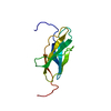
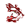
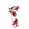
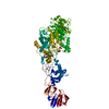
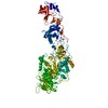
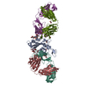
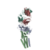
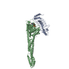
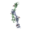
Entry Database : PDB / ID : 4y61Title Crystal structure of the complex between Slitrk2 LRR1 and PTP delta Ig1-Fn1 Receptor-type tyrosine-protein phosphatase delta SLIT and NTRK-like protein 2 Keywords / / Function / homology Function Domain/homology Component
/ / / / / / / / / / / / / / / / / / / / / / / / / / / / / / / / / / / / / / / / / / / / / / / / / / / / / / / / / / / / / / / / / / / / / / / / / / / / / / / / / / / / / / / / / / / / / / / / Biological species Mus musculus (house mouse)Method / / / Resolution : 3.358 Å Authors Yamgata, A. / Sato, Y. / Goto-Ito, S. / Uemura, T. / Maeda, A. / Shiroshima, T. / Yoshida, T. / Fukai, S. Funding support Organization Grant number Country
Journal : Sci Rep / Year : 2015Title : Structure of Slitrk2-PTP delta complex reveals mechanisms for splicing-dependent trans-synaptic adhesion.Authors : Yamagata, A. / Sato, Y. / Goto-Ito, S. / Uemura, T. / Maeda, A. / Shiroshima, T. / Yoshida, T. / Fukai, S. History Deposition Feb 12, 2015 Deposition site / Processing site Revision 1.0 Jun 3, 2015 Provider / Type Revision 1.1 Oct 4, 2017 Group / Derived calculations / Source and taxonomyCategory diffrn_detector / diffrn_source ... diffrn_detector / diffrn_source / entity_src_gen / pdbx_struct_assembly / pdbx_struct_assembly_gen / pdbx_struct_assembly_prop / pdbx_struct_oper_list Item _diffrn_detector.detector / _diffrn_source.pdbx_synchrotron_site ... _diffrn_detector.detector / _diffrn_source.pdbx_synchrotron_site / _entity_src_gen.pdbx_alt_source_flag / _pdbx_struct_assembly.oligomeric_details / _pdbx_struct_assembly_gen.asym_id_list / _pdbx_struct_assembly_prop.type / _pdbx_struct_assembly_prop.value / _pdbx_struct_oper_list.symmetry_operation Revision 1.2 Jul 29, 2020 Group / Derived calculations / Structure summaryCategory chem_comp / entity ... chem_comp / entity / pdbx_chem_comp_identifier / pdbx_entity_nonpoly / struct_conn / struct_site / struct_site_gen Item _chem_comp.name / _chem_comp.type ... _chem_comp.name / _chem_comp.type / _entity.pdbx_description / _pdbx_entity_nonpoly.name / _struct_conn.pdbx_role Description / Provider / Type Revision 1.3 Nov 8, 2023 Group Data collection / Database references ... Data collection / Database references / Refinement description / Structure summary Category chem_comp / chem_comp_atom ... chem_comp / chem_comp_atom / chem_comp_bond / database_2 / pdbx_initial_refinement_model Item / _database_2.pdbx_DOI / _database_2.pdbx_database_accessionRevision 1.4 Nov 13, 2024 Group / Category / pdbx_modification_feature
Show all Show less
 Movie
Movie Controller

 Yorodumi
 Open data
 Basic information
 Components
 Keywords
 Function and homology information
 X-RAY DIFFRACTION /
  Authors
 Japan, 1items
  Citation
 Structure visualization
 Downloads & links
 Other downloads
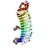


 PDBj
PDBj


 Assembly
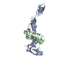
 Homo sapiens (human) / References: UniProt: Q64487, protein-tyrosine-phosphatase
 Type: D-saccharide, beta linking / Mass: 221.208 Da / Num. of mol.: 4 / Source method: obtained synthetically / Formula: C8H15NO6
 Sample preparation
 Processing