 Movie
Movie Controller

[English] 日本語
 Yorodumi
- PDB-3kll: Crystal structure of Lactobacillus reuteri N-terminally truncated... -
+ Open data

- Basic information
| Entry | Database: PDB / ID: 3kll | |||||||||
|---|---|---|---|---|---|---|---|---|---|---|
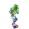
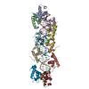
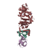
| Title | Crystal structure of Lactobacillus reuteri N-terminally truncated glucansucrase GTF180-maltose complex | |||||||||
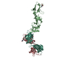
 Components Components | Glucansucrase | |||||||||
 Keywords Keywords | TRANSFERASE / glucansucrase-maltose complex / multidomain protein / Glycosyltransferase | |||||||||
| Function / homology |  Function and homology information Function and homology informationdextransucrase activity / dextransucrase / glucan biosynthetic process / glucosyltransferase activity / metal ion binding Similarity search - Function | |||||||||
| Biological species |  Lactobacillus reuteri (bacteria) Lactobacillus reuteri (bacteria) | |||||||||
| Method |  X-RAY DIFFRACTION / SYNCHROTRON / MOLECULAR REPLACEMENT / molecular replacement / Resolution: 2 Å X-RAY DIFFRACTION / SYNCHROTRON / MOLECULAR REPLACEMENT / molecular replacement / Resolution: 2 Å | |||||||||
 Authors Authors | Vujicic-Zagar, A. / Pijning, T. / Kralj, S. / Eeuwema, W. / Dijkhuizen, L. / Dijkstra, B.W. | |||||||||
 Citation Citation | Journal: Proc.Natl.Acad.Sci.USA / Year: 2010 Title: Crystal structure of a 117 kDa glucansucrase fragment provides insight into evolution and product specificity of GH70 enzymes Authors: Vujicic-Zagar, A. / Pijning, T. / Kralj, S. / Lopez, C.A. / Eeuwema, W. / Dijkhuizen, L. / Dijkstra, B.W. | |||||||||
| History |
|
- Structure visualization
| Structure viewer | Molecule: MolmilJmol/JSmol |
|---|
- Downloads & links
-Download
| PDBx/mmCIF format | 3kll.cif.gz | 237.9 KB | Display | PDBx/mmCIF format |
|---|---|---|---|---|
| PDB format | pdb3kll.ent.gz | 182.2 KB | Display | PDB format |
| PDBx/mmJSON format | 3kll.json.gz | Tree view | PDBx/mmJSON format | |
| Others |  Other downloads Other downloads |
-Validation report
| Arichive directory | https://data.pdbj.org/pub/pdb/validation_reports/kl/3kllftp://data.pdbj.org/pub/pdb/validation_reports/kl/3kll | HTTPS FTP |
|---|
-Related structure data
| Related structure data |  3hz3C  3klkSC  3hq3 C: citing same article ( S: Starting model for refinement |
|---|---|
| Similar structure data |


-Links
 PDBj
PDBj


- Assembly
| Deposited unit | 
| ||||||||
|---|---|---|---|---|---|---|---|---|---|
| 1 |
| ||||||||
| Unit cell |
|
-Components
| #1: Protein | Mass: 117767.336 Da / Num. of mol.: 1 Fragment: N-terminally truncated GTF180, UNP residues 742-1772 Source method: isolated from a genetically manipulated source Source: (gene. exp.) Lactobacillus reuteri (bacteria) / Strain: 180 / Gene: gtf180 / Plasmid: pET15b / Production host: | ||||||||||
|---|---|---|---|---|---|---|---|---|---|---|---|
| #2: Polysaccharide | alpha-D-glucopyranose-(1-4)-alpha-D-glucopyranose / alpha-maltose   Source method: isolated from a genetically manipulated source Details: oligosaccharide / References: alpha-maltose #3: Chemical | ChemComp-CA / |   Mass: 40.078 Da / Num. of mol.: 1 / Source method: obtained synthetically / Formula: Ca Mass: 40.078 Da / Num. of mol.: 1 / Source method: obtained synthetically / Formula: Ca#4: Chemical | ChemComp-GOL /   Mass: 92.094 Da / Num. of mol.: 4 / Source method: obtained synthetically / Formula: C3H8O3 Mass: 92.094 Da / Num. of mol.: 4 / Source method: obtained synthetically / Formula: C3H8O3#5: Water | ChemComp-HOH / |  Mass: 18.015 Da / Num. of mol.: 758 / Source method: isolated from a natural source / Formula: H2O Mass: 18.015 Da / Num. of mol.: 758 / Source method: isolated from a natural source / Formula: H2OHas protein modification | Y | Sequence details | THE CONFLICT F1674L MAY DUE TO PCR ERROR. | |
-Experimental details
-Experiment
| Experiment | Method: X-RAY DIFFRACTION / Number of used crystals: 1 |
|---|
- Sample preparation
| Crystal | Density Matthews: 2.52 Å3/Da / Density % sol: 51.1 % / Mosaicity: 0 ° |
|---|---|
| Crystal grow | Temperature: 293 K / Method: vapor diffusion, hanging drop / pH: 6 Details: PEG 3350, BIS-TRIS propane buffer, CaCl2, pH 6.0, vapor diffusion, hanging drop, temperature 293K |
-Data collection
| Diffraction | Mean temperature: 100 K |
|---|---|
| Diffraction source | Source: SYNCHROTRON / Site: EMBL/DESY, HAMBURG  / Beamline: BW7A / Wavelength: 0.976 Å / Beamline: BW7A / Wavelength: 0.976 Å |
| Detector | Type: MAR CCD 165 mm / Detector: CCD / Date: Feb 24, 2006 |
| Radiation | Protocol: SINGLE WAVELENGTH / Monochromatic (M) / Laue (L): M / Scattering type: x-ray |
| Radiation wavelength | Wavelength: 0.976 Å / Relative weight: 1 |
| Reflection | Resolution: 1.997→38.747 Å / Num. obs: 75992 / % possible obs: 97.5 % / Redundancy: 2.4 % / Rsym value: 0.053 |
| Reflection shell | Resolution: 2→2.1 Å / Redundancy: 2.4 % / Mean I/σ(I) obs: 4.3 / Num. unique all: 10726 / Rsym value: 0.171 / % possible all: 94 |
-Phasing
| Phasing | Method: molecular replacement |
|---|
- Processing
| Software |
| |||||||||||||||||||||||||||||||||||||||||||||||||||||||||||||||||
|---|---|---|---|---|---|---|---|---|---|---|---|---|---|---|---|---|---|---|---|---|---|---|---|---|---|---|---|---|---|---|---|---|---|---|---|---|---|---|---|---|---|---|---|---|---|---|---|---|---|---|---|---|---|---|---|---|---|---|---|---|---|---|---|---|---|---|
| Refinement | Method to determine structure: MOLECULAR REPLACEMENT Starting model: PDB ENTRY 3KLK Resolution: 2→20 Å / Cor.coef. Fo:Fc: 0.962 / Cor.coef. Fo:Fc free: 0.945 / Occupancy max: 1 / Occupancy min: 0.8 / SU B: 3.576 / SU ML: 0.099 / Cross valid method: THROUGHOUT / σ(F): 0 / ESU R: 0.164 / ESU R Free: 0.143 / Stereochemistry target values: MAXIMUM LIKELIHOOD Details: HYDROGENS HAVE BEEN ADDED IN THE RIDING POSITIONS U VALUES: REFINED INDIVIDUALLY
| |||||||||||||||||||||||||||||||||||||||||||||||||||||||||||||||||
| Solvent computation | Ion probe radii: 0.8 Å / Shrinkage radii: 0.8 Å / VDW probe radii: 1.4 Å / Solvent model: BABINET MODEL WITH MASK | |||||||||||||||||||||||||||||||||||||||||||||||||||||||||||||||||
| Displacement parameters | Biso max: 233.6 Å2 / Biso mean: 29.568 Å2 / Biso min: 13.35 Å2
| |||||||||||||||||||||||||||||||||||||||||||||||||||||||||||||||||
| Refinement step | Cycle: LAST / Resolution: 2→20 Å
| |||||||||||||||||||||||||||||||||||||||||||||||||||||||||||||||||
| Refine LS restraints |
| |||||||||||||||||||||||||||||||||||||||||||||||||||||||||||||||||
| LS refinement shell | Resolution: 2→2.051 Å / Total num. of bins used: 20
|
