 Movie
Movie Controller

[English] 日本語
 Yorodumi
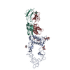
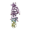
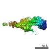
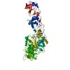
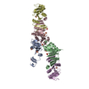
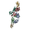
- PDB-7jic: Structure of human CD19-CD81 co-receptor complex bound to coltuxi... -
+ Open data

- Basic information
| Entry | Database: PDB / ID: 7jic | ||||||||||||
|---|---|---|---|---|---|---|---|---|---|---|---|---|---|
| Title | Structure of human CD19-CD81 co-receptor complex bound to coltuximab Fab fragment | ||||||||||||
 Components Components |
| ||||||||||||
 Keywords Keywords | IMMUNE SYSTEM / tetraspanin / Fab / complex | ||||||||||||
| Function / homology |  Function and homology information Function and homology informationregulation of B cell activation / positive regulation of adaptive immune memory response / positive regulation of protein catabolic process in the vacuole / antigen receptor-mediated signaling pathway / CD4-positive, alpha-beta T cell costimulation / positive regulation of B cell receptor signaling pathway / osteoclast fusion / B-1 B cell differentiation / myoblast fusion involved in skeletal muscle regeneration / positive regulation of T cell activation via T cell receptor contact with antigen bound to MHC molecule on antigen presenting cell ...regulation of B cell activation / positive regulation of adaptive immune memory response / positive regulation of protein catabolic process in the vacuole / antigen receptor-mediated signaling pathway / CD4-positive, alpha-beta T cell costimulation / positive regulation of B cell receptor signaling pathway / osteoclast fusion / B-1 B cell differentiation / myoblast fusion involved in skeletal muscle regeneration / positive regulation of T cell activation via T cell receptor contact with antigen bound to MHC molecule on antigen presenting cell / regulation of B cell receptor signaling pathway / positive regulation of inflammatory response to antigenic stimulus / regulation of macrophage migration / macrophage fusion / immunological synapse formation / tetraspanin-enriched microdomain / positive regulation of T-helper 2 cell cytokine production / B cell proliferation involved in immune response / transferrin receptor binding / humoral immune response mediated by circulating immunoglobulin / positive regulation of protein exit from endoplasmic reticulum / protein localization to lysosome / MHC class II protein binding / positive regulation of CD4-positive, alpha-beta T cell proliferation / positive regulation of T cell receptor signaling pathway / cholesterol binding / immunoglobulin mediated immune response / B cell proliferation / immunological synapse / cellular response to low-density lipoprotein particle stimulus / positive regulation of receptor clustering / positive regulation of B cell proliferation / Regulation of Complement cascade / basal plasma membrane / Antigen activates B Cell Receptor (BCR) leading to generation of second messengers / positive regulation of release of sequestered calcium ion into cytosol / B cell receptor signaling pathway / protein localization to plasma membrane / regulation of protein stability / receptor internalization / integrin binding / Immunoregulatory interactions between a Lymphoid and a non-Lymphoid cell / Constitutive Signaling by Aberrant PI3K in Cancer / MHC class II protein complex binding / PIP3 activates AKT signaling / PI5P, PP2A and IER3 Regulate PI3K/AKT Signaling / virus receptor activity / vesicle / basolateral plasma membrane / positive regulation of MAPK cascade / positive regulation of phosphatidylinositol 3-kinase/protein kinase B signal transduction / membrane raft / external side of plasma membrane / focal adhesion / positive regulation of transcription by RNA polymerase II / protein-containing complex / extracellular exosome / membrane / plasma membrane / cytosol Similarity search - Function | ||||||||||||
| Biological species |  Homo sapiens (human) Homo sapiens (human) | ||||||||||||
| Method | ELECTRON MICROSCOPY / single particle reconstruction / cryo EM / Resolution: 3.8 Å | ||||||||||||
 Authors Authors | Susa, K.J. / Rawson, S. / Kruse, A.C. / Blacklow, S.C. | ||||||||||||
| Funding support |  United States, 3items United States, 3items
| ||||||||||||
 Citation Citation | Journal: Science / Year: 2021 Title: Cryo-EM structure of the B cell co-receptor CD19 bound to the tetraspanin CD81. Authors: Katherine J Susa / Shaun Rawson / Andrew C Kruse / Stephen C Blacklow / Abstract: Signaling through the CD19-CD81 co-receptor complex, in combination with the B cell receptor, is a critical determinant of B cell development and activation. It is unknown how CD81 engages CD19 to ...Signaling through the CD19-CD81 co-receptor complex, in combination with the B cell receptor, is a critical determinant of B cell development and activation. It is unknown how CD81 engages CD19 to enable co-receptor function. Here, we report a 3.8-angstrom structure of the CD19-CD81 complex bound to a therapeutic antigen-binding fragment, determined by cryo-electron microscopy (cryo-EM). The structure includes both the extracellular domains and the transmembrane helices of the complex, revealing a contact interface between the ectodomains that drives complex formation. Upon binding to CD19, CD81 opens its ectodomain to expose a hydrophobic CD19-binding surface and reorganizes its transmembrane helices to occlude a cholesterol binding pocket present in the apoprotein. Our data reveal the structural basis for CD19-CD81 complex assembly, providing a foundation for rational design of therapies for B cell dysfunction. | ||||||||||||
| History |
|
- Structure visualization
| Movie |
Movie viewer |
|---|---|
| Structure viewer | Molecule: MolmilJmol/JSmol |
- Downloads & links
-Download
| PDBx/mmCIF format | 7jic.cif.gz | 159.9 KB | Display | PDBx/mmCIF format |
|---|---|---|---|---|
| PDB format | pdb7jic.ent.gz | 115 KB | Display | PDB format |
| PDBx/mmJSON format | 7jic.json.gz | Tree view | PDBx/mmJSON format | |
| Others |  Other downloads Other downloads |
-Validation report
| Arichive directory | https://data.pdbj.org/pub/pdb/validation_reports/ji/7jicftp://data.pdbj.org/pub/pdb/validation_reports/ji/7jic | HTTPS FTP |
|---|
-Related structure data
| Related structure data |  22344MC M: map data used to model this data C: citing same article ( |
|---|---|
| Similar structure data |


-Links
 PDBj
PDBj


- Assembly
| Deposited unit | 
|
|---|---|
| 1 |
|
-Components
| #1: Protein | Mass: 36031.578 Da / Num. of mol.: 1 Source method: isolated from a genetically manipulated source Source: (gene. exp.) Homo sapiens (human) / Gene: CD19 / Plasmid: pcDNA3.1 / Cell line (production host): Expi293F / Production host: Homo sapiens (human) / References: UniProt: P15391 |
|---|---|
| #2: Protein | Mass: 26444.758 Da / Num. of mol.: 1 Source method: isolated from a genetically manipulated source Source: (gene. exp.) Homo sapiens (human) / Gene: CD81, TAPA1, TSPAN28 / Plasmid: pcDNA3.1 / Cell line (production host): Expi293F / Production host: Homo sapiens (human) / References: UniProt: P60033 |
| #3: Antibody | Mass: 24828.639 Da / Num. of mol.: 1 Source method: isolated from a genetically manipulated source Source: (gene. exp.) Homo sapiens (human) |
| #4: Antibody | Mass: 23211.758 Da / Num. of mol.: 1 Source method: isolated from a genetically manipulated source Source: (gene. exp.) Homo sapiens (human) |
| Has protein modification | Y |
-Experimental details
-Experiment
| Experiment | Method: ELECTRON MICROSCOPY |
|---|---|
| EM experiment | Aggregation state: PARTICLE / 3D reconstruction method: single particle reconstruction |
- Sample preparation
| Component |
| |||||||||||||||||||||||||
|---|---|---|---|---|---|---|---|---|---|---|---|---|---|---|---|---|---|---|---|---|---|---|---|---|---|---|
| Molecular weight | Value: 0.110 MDa / Experimental value: NO | |||||||||||||||||||||||||
| Source (natural) |
| |||||||||||||||||||||||||
| Source (recombinant) |
| |||||||||||||||||||||||||
| Buffer solution | pH: 7.4 | |||||||||||||||||||||||||
| Buffer component |
| |||||||||||||||||||||||||
| Specimen | Conc.: 1.8 mg/ml / Embedding applied: NO / Shadowing applied: NO / Staining applied: NO / Vitrification applied: YES | |||||||||||||||||||||||||
| Specimen support | Grid material: COPPER / Grid mesh size: 400 divisions/in. / Grid type: Quantifoil | |||||||||||||||||||||||||
| Vitrification | Instrument: FEI VITROBOT MARK IV / Cryogen name: ETHANE / Humidity: 100 % / Chamber temperature: 295 K Details: Blot for 4.5-5.5 seconds with a blot force of 15-16. |
- Electron microscopy imaging
| Experimental equipment |  Model: Titan Krios / Image courtesy: FEI Company |
|---|---|
| Microscopy | Model: FEI TITAN KRIOS |
| Electron gun | Electron source:  FIELD EMISSION GUN / Accelerating voltage: 300 kV / Illumination mode: FLOOD BEAM FIELD EMISSION GUN / Accelerating voltage: 300 kV / Illumination mode: FLOOD BEAM |
| Electron lens | Mode: BRIGHT FIELD / Nominal magnification: 105000 X / Nominal defocus max: 2500 nm / Nominal defocus min: 1000 nm / Cs: 2.7 mm |
| Specimen holder | Specimen holder model: FEI TITAN KRIOS AUTOGRID HOLDER |
| Image recording | Electron dose: 55 e/Å2 / Film or detector model: GATAN K3 BIOQUANTUM (6k x 4k) / Num. of grids imaged: 6 |
- Processing
| Software |
| |||||||||||||||||||||||||
|---|---|---|---|---|---|---|---|---|---|---|---|---|---|---|---|---|---|---|---|---|---|---|---|---|---|---|
| EM software |
| |||||||||||||||||||||||||
| CTF correction | Type: PHASE FLIPPING AND AMPLITUDE CORRECTION | |||||||||||||||||||||||||
| Particle selection | Num. of particles selected: 2798945 | |||||||||||||||||||||||||
| Symmetry | Point symmetry: C1 (asymmetric) | |||||||||||||||||||||||||
| 3D reconstruction | Resolution: 3.8 Å / Resolution method: FSC 0.143 CUT-OFF / Num. of particles: 244583 / Num. of class averages: 1 / Symmetry type: POINT | |||||||||||||||||||||||||
| Atomic model building | 3D fitting-ID: 1 / Source name: PDB / Type: experimental model
| |||||||||||||||||||||||||
| Refinement | Cross valid method: NONE Stereochemistry target values: GeoStd + Monomer Library + CDL v1.2 | |||||||||||||||||||||||||
| Displacement parameters | Biso mean: 114.72 Å2 | |||||||||||||||||||||||||
| Refine LS restraints |
|


