- EMDB-22344: Structure of human CD19-CD81 co-receptor complex bound to coltuxi... -
+
Open data
ID or keywords:
Loading...
-
Basic information
Entry
Database: EMDB / ID: EMD-22344
Title
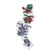
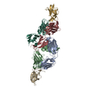
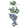
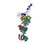
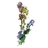
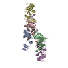
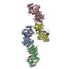
Structure of human CD19-CD81 co-receptor complex bound to coltuximab Fab fragment
Map data
Sample
Complex: Complex of B cell co-receptor CD19 bound to the tetraspanin CD81 and coltuximab Fab fragment
Complex: B cell co-receptor CD19 bound to the tetraspanin CD81
Protein or peptide: B-lymphocyte antigen CD19
Protein or peptide: CD81 antigen
Complex: coltuximab Fab fragment
Protein or peptide: Coltuximab Heavy Chain
Protein or peptide: Coltuximab Light Chain
Keywords
tetraspanin / Fab / complex / IMMUNE SYSTEM
Function / homology
Function and homology information
regulation of B cell activation / positive regulation of adaptive immune memory response / positive regulation of protein catabolic process in the vacuole / antigen receptor-mediated signaling pathway / CD4-positive, alpha-beta T cell costimulation / positive regulation of B cell receptor signaling pathway / osteoclast fusion / B-1 B cell differentiation / myoblast fusion involved in skeletal muscle regeneration / positive regulation of T cell activation via T cell receptor contact with antigen bound to MHC molecule on antigen presenting cell ...regulation of B cell activation / positive regulation of adaptive immune memory response / positive regulation of protein catabolic process in the vacuole / antigen receptor-mediated signaling pathway / CD4-positive, alpha-beta T cell costimulation / positive regulation of B cell receptor signaling pathway / osteoclast fusion / B-1 B cell differentiation / myoblast fusion involved in skeletal muscle regeneration / positive regulation of T cell activation via T cell receptor contact with antigen bound to MHC molecule on antigen presenting cell / regulation of B cell receptor signaling pathway / positive regulation of inflammatory response to antigenic stimulus / regulation of macrophage migration / macrophage fusion / immunological synapse formation / tetraspanin-enriched microdomain / positive regulation of T-helper 2 cell cytokine production / B cell proliferation involved in immune response / transferrin receptor binding / humoral immune response mediated by circulating immunoglobulin / positive regulation of protein exit from endoplasmic reticulum / protein localization to lysosome / MHC class II protein binding / positive regulation of CD4-positive, alpha-beta T cell proliferation / positive regulation of T cell receptor signaling pathway / cholesterol binding / immunoglobulin mediated immune response / B cell proliferation / immunological synapse / cellular response to low-density lipoprotein particle stimulus / positive regulation of receptor clustering / positive regulation of B cell proliferation / Regulation of Complement cascade / basal plasma membrane / Antigen activates B Cell Receptor (BCR) leading to generation of second messengers / positive regulation of release of sequestered calcium ion into cytosol / B cell receptor signaling pathway / protein localization to plasma membrane / regulation of protein stability / receptor internalization / integrin binding / Immunoregulatory interactions between a Lymphoid and a non-Lymphoid cell / Constitutive Signaling by Aberrant PI3K in Cancer / MHC class II protein complex binding / PIP3 activates AKT signaling / PI5P, PP2A and IER3 Regulate PI3K/AKT Signaling / virus receptor activity / vesicle / basolateral plasma membrane / positive regulation of MAPK cascade / positive regulation of phosphatidylinositol 3-kinase/protein kinase B signal transduction / membrane raft / external side of plasma membrane / focal adhesion / positive regulation of transcription by RNA polymerase II / protein-containing complex / extracellular exosome / membrane / plasma membrane / cytosol Similarity search - Function
National Institutes of Health/National Heart, Lung, and Blood Institute (NIH/NHLBI)
HL 147459
United States
National Institutes of Health/National Cancer Institute (NIH/NCI)
R35 CA220340
United States
National Institutes of Health/Office of the Director
DP5 OD021345
United States
Citation
Journal: Science / Year: 2021 Title: Cryo-EM structure of the B cell co-receptor CD19 bound to the tetraspanin CD81. Authors: Katherine J Susa / Shaun Rawson / Andrew C Kruse / Stephen C Blacklow / Abstract: Signaling through the CD19-CD81 co-receptor complex, in combination with the B cell receptor, is a critical determinant of B cell development and activation. It is unknown how CD81 engages CD19 to ...Signaling through the CD19-CD81 co-receptor complex, in combination with the B cell receptor, is a critical determinant of B cell development and activation. It is unknown how CD81 engages CD19 to enable co-receptor function. Here, we report a 3.8-angstrom structure of the CD19-CD81 complex bound to a therapeutic antigen-binding fragment, determined by cryo-electron microscopy (cryo-EM). The structure includes both the extracellular domains and the transmembrane helices of the complex, revealing a contact interface between the ectodomains that drives complex formation. Upon binding to CD19, CD81 opens its ectodomain to expose a hydrophobic CD19-binding surface and reorganizes its transmembrane helices to occlude a cholesterol binding pocket present in the apoprotein. Our data reveal the structural basis for CD19-CD81 complex assembly, providing a foundation for rational design of therapies for B cell dysfunction.
History
Deposition
Jul 23, 2020
-
Header (metadata) release
Jan 20, 2021
-
Map release
Jan 20, 2021
-
Update
Oct 30, 2024
-
Current status
Oct 30, 2024
Processing site: RCSB / Status: Released
-
Structure visualization
Movie
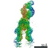
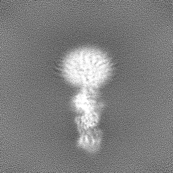
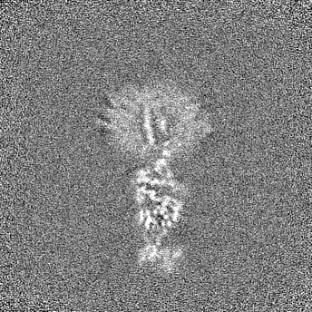
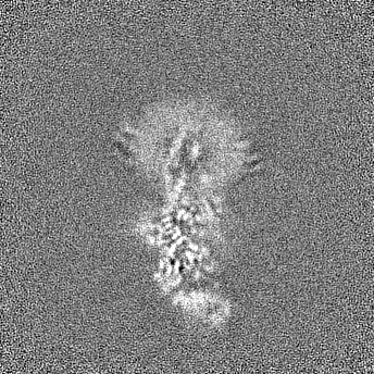
Surface view with section colored by density value
Entire : Complex of B cell co-receptor CD19 bound to the tetraspanin CD81 ...
Entire
Name: Complex of B cell co-receptor CD19 bound to the tetraspanin CD81 and coltuximab Fab fragment
Components
Complex: Complex of B cell co-receptor CD19 bound to the tetraspanin CD81 and coltuximab Fab fragment
Complex: B cell co-receptor CD19 bound to the tetraspanin CD81
Protein or peptide: B-lymphocyte antigen CD19
Protein or peptide: CD81 antigen
Complex: coltuximab Fab fragment
Protein or peptide: Coltuximab Heavy Chain
Protein or peptide: Coltuximab Light Chain
-
Supramolecule #1: Complex of B cell co-receptor CD19 bound to the tetraspanin CD81 ...
Supramolecule
Name: Complex of B cell co-receptor CD19 bound to the tetraspanin CD81 and coltuximab Fab fragment type: complex / ID: 1 / Parent: 0 / Macromolecule list: all
Molecular weight
Theoretical: 110 KDa
-
Supramolecule #2: B cell co-receptor CD19 bound to the tetraspanin CD81
Supramolecule
Name: B cell co-receptor CD19 bound to the tetraspanin CD81 / type: complex / ID: 2 / Parent: 1 / Macromolecule list: #1-#2
Source (natural)
Organism: Homo sapiens (human)
-
Supramolecule #3: coltuximab Fab fragment
Supramolecule
Name: coltuximab Fab fragment / type: complex / ID: 3 / Parent: 1 / Macromolecule list: #3-#4
Source (natural)
Organism: Mus musculus (house mouse)
-
Macromolecule #1: B-lymphocyte antigen CD19
Macromolecule
Name: B-lymphocyte antigen CD19 / type: protein_or_peptide / ID: 1 / Number of copies: 1 / Enantiomer: LEVO
Cryogen name: ETHANE / Chamber humidity: 100 % / Chamber temperature: 295 K / Instrument: FEI VITROBOT MARK IV Details: Blot for 4.5-5.5 seconds with a blot force of 15-16..
-
Electron microscopy
Microscope
FEI TITAN KRIOS
Image recording
Film or detector model: GATAN K3 BIOQUANTUM (6k x 4k) / Number grids imaged: 6 / Average electron dose: 55.0 e/Å2
Electron beam
Acceleration voltage: 300 kV / Electron source: FIELD EMISSION GUN
In the structure databanks used in Yorodumi, some data are registered as the other names, "COVID-19 virus" and "2019-nCoV". Here are the details of the virus and the list of structure data.
Jan 31, 2019. EMDB accession codes are about to change! (news from PDBe EMDB page)
EMDB accession codes are about to change! (news from PDBe EMDB page)
The allocation of 4 digits for EMDB accession codes will soon come to an end. Whilst these codes will remain in use, new EMDB accession codes will include an additional digit and will expand incrementally as the available range of codes is exhausted. The current 4-digit format prefixed with “EMD-” (i.e. EMD-XXXX) will advance to a 5-digit format (i.e. EMD-XXXXX), and so on. It is currently estimated that the 4-digit codes will be depleted around Spring 2019, at which point the 5-digit format will come into force.
The EM Navigator/Yorodumi systems omit the EMD- prefix.
Related info.:Q: What is EMD? / ID/Accession-code notation in Yorodumi/EM Navigator
Yorodumi is a browser for structure data from EMDB, PDB, SASBDB, etc.
This page is also the successor to EM Navigator detail page, and also detail information page/front-end page for Omokage search.
The word "yorodu" (or yorozu) is an old Japanese word meaning "ten thousand". "mi" (miru) is to see.
Related info.:EMDB / PDB / SASBDB / Comparison of 3 databanks / Yorodumi Search / Aug 31, 2016. New EM Navigator & Yorodumi / Yorodumi Papers / Jmol/JSmol / Function and homology information / Changes in new EM Navigator and Yorodumi
 Movie
Movie Controller

 Yorodumi
 Open data
 Basic information
 Map data
 Sample
 Keywords
 Function and homology information
 Homo sapiens (human) /
 Authors
 United States, 3 items
  Citation
 Structure visualization
 Downloads & links
 emd_22344.png
 http://ftp.pdbj.org/pub/emdb/structures/EMD-22344


 Z (Sec.)
 Y (Row.)
 X (Col.)


 Sample components
 Processing
 Electron microscopy
 FIELD EMISSION GUN


