 Movie
Movie Controller


+ Open data

- Basic information
| Entry | Database: PDB / ID: 6lng | ||||||
|---|---|---|---|---|---|---|---|
| Title | Rapid crystallization of streptavidin using charged peptides | ||||||
 Components Components | Streptavidin | ||||||
 Keywords Keywords | PEPTIDE BINDING PROTEIN / BIOTIN-BINDING PROTEIN | ||||||
| Function / homology |  Function and homology information Function and homology information | ||||||
| Biological species |  Streptomyces avidinii (bacteria) Streptomyces avidinii (bacteria) | ||||||
| Method |  X-RAY DIFFRACTION / SYNCHROTRON / MOLECULAR REPLACEMENT / Resolution: 1.80000150972 Å X-RAY DIFFRACTION / SYNCHROTRON / MOLECULAR REPLACEMENT / Resolution: 1.80000150972 Å | ||||||
 Authors Authors | Minamihata, K. / Tsukamoto, K. / Adachi, M. / Shimizu, R. / Mishina, M. / Kuroki, R. / Nagamune, T. | ||||||
 Citation Citation | Journal: Chem.Commun.(Camb.) / Year: 2020 Title: Genetically fused charged peptides induce rapid crystallization of proteins. Authors: Minamihata, K. / Tsukamoto, K. / Adachi, M. / Shimizu, R. / Mishina, M. / Kuroki, R. / Nagamune, T. | ||||||
| History |
|
- Structure visualization
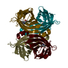
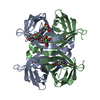
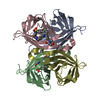
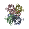
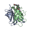
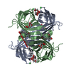
| Structure viewer | Molecule: MolmilJmol/JSmol |
|---|
- Downloads & links
-Download
| PDBx/mmCIF format | 6lng.cif.gz | 198.9 KB | Display | PDBx/mmCIF format |
|---|---|---|---|---|
| PDB format | pdb6lng.ent.gz | 128.3 KB | Display | PDB format |
| PDBx/mmJSON format | 6lng.json.gz | Tree view | PDBx/mmJSON format | |
| Others |  Other downloads Other downloads |
-Validation report
| Arichive directory | https://data.pdbj.org/pub/pdb/validation_reports/ln/6lngftp://data.pdbj.org/pub/pdb/validation_reports/ln/6lng | HTTPS FTP |
|---|
-Related structure data
| Related structure data |  3ry1S S: Starting model for refinement |
|---|---|
| Similar structure data |


-Links
 PDBj
PDBj- Assembly
| Deposited unit | 
| ||||||||||
|---|---|---|---|---|---|---|---|---|---|---|---|
| 1 | 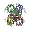
| ||||||||||
| 2 | 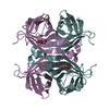
| ||||||||||
| Unit cell |
| ||||||||||
| Components on special symmetry positions |
|
-Components
| #1: Protein | Mass: 13081.144 Da / Num. of mol.: 6 Source method: isolated from a genetically manipulated source Details: Mixture of Two kinds of proteins, which have RRRRRRY or DDDDDDY attached at C-terminal respectively, was used in this study. Source: (gene. exp.) Streptomyces avidinii (bacteria) / Production host: #2: Chemical | ChemComp-GOL /   Mass: 92.094 Da / Num. of mol.: 6 / Source method: obtained synthetically / Formula: C3H8O3 / Feature type: SUBJECT OF INVESTIGATION Mass: 92.094 Da / Num. of mol.: 6 / Source method: obtained synthetically / Formula: C3H8O3 / Feature type: SUBJECT OF INVESTIGATION#3: Water | ChemComp-HOH / |  Mass: 18.015 Da / Num. of mol.: 712 / Source method: isolated from a natural source / Formula: H2O Mass: 18.015 Da / Num. of mol.: 712 / Source method: isolated from a natural source / Formula: H2OHas ligand of interest | Y | |
|---|
-Experimental details
-Experiment
| Experiment | Method: X-RAY DIFFRACTION / Number of used crystals: 1 |
|---|
- Sample preparation
| Crystal | Density Matthews: 2.74 Å3/Da / Density % sol: 55.05 % |
|---|---|
| Crystal grow | Temperature: 298 K / Method: batch mode Details: 0.2 M ammonium sulfate, 0.1 M MES pH 6.5, 30% w/v polyethylene glycol monomethyl ether 5000 |
-Data collection
| Diffraction | Mean temperature: 100 K / Serial crystal experiment: N |
|---|---|
| Diffraction source | Source: SYNCHROTRON / Site: SPring-8  / Beamline: BL38B1 / Wavelength: 1 Å / Beamline: BL38B1 / Wavelength: 1 Å |
| Detector | Type: ADSC QUANTUM 315r / Detector: CCD / Date: Nov 22, 2014 |
| Radiation | Protocol: SINGLE WAVELENGTH / Monochromatic (M) / Laue (L): M / Scattering type: x-ray |
| Radiation wavelength | Wavelength: 1 Å / Relative weight: 1 |
| Reflection | Resolution: 1.8→37.1 Å / Num. obs: 78148 / % possible obs: 99.7 % / Redundancy: 3.67 % / Biso Wilson estimate: 16.1760765111 Å2 / Rmerge(I) obs: 0.064 / Net I/σ(I): 13.91 |
| Reflection shell | Resolution: 1.8→1.85 Å / Rmerge(I) obs: 0.39 / Mean I/σ(I) obs: 3.1 / Num. unique obs: 5742 |
- Processing
| Software |
| |||||||||||||||||||||||||||||||||||||||||||||||||||||||||||||||||||||||||||||||||||||||||||||||||||||||||||||||||||||||||||||||||||||||||||||||||||||||||||||||||||||||||||||||||||||||||||||||||||||||||||
|---|---|---|---|---|---|---|---|---|---|---|---|---|---|---|---|---|---|---|---|---|---|---|---|---|---|---|---|---|---|---|---|---|---|---|---|---|---|---|---|---|---|---|---|---|---|---|---|---|---|---|---|---|---|---|---|---|---|---|---|---|---|---|---|---|---|---|---|---|---|---|---|---|---|---|---|---|---|---|---|---|---|---|---|---|---|---|---|---|---|---|---|---|---|---|---|---|---|---|---|---|---|---|---|---|---|---|---|---|---|---|---|---|---|---|---|---|---|---|---|---|---|---|---|---|---|---|---|---|---|---|---|---|---|---|---|---|---|---|---|---|---|---|---|---|---|---|---|---|---|---|---|---|---|---|---|---|---|---|---|---|---|---|---|---|---|---|---|---|---|---|---|---|---|---|---|---|---|---|---|---|---|---|---|---|---|---|---|---|---|---|---|---|---|---|---|---|---|---|---|---|---|---|---|---|
| Refinement | Method to determine structure: MOLECULAR REPLACEMENT Starting model: 3RY1 Resolution: 1.80000150972→37.0878676876 Å / SU ML: 0.181418679282 / Cross valid method: FREE R-VALUE / σ(F): 1.3434970804 / Phase error: 22.4923699741
| |||||||||||||||||||||||||||||||||||||||||||||||||||||||||||||||||||||||||||||||||||||||||||||||||||||||||||||||||||||||||||||||||||||||||||||||||||||||||||||||||||||||||||||||||||||||||||||||||||||||||||
| Solvent computation | Shrinkage radii: 0.9 Å / VDW probe radii: 1.11 Å / Solvent model: FLAT BULK SOLVENT MODEL | |||||||||||||||||||||||||||||||||||||||||||||||||||||||||||||||||||||||||||||||||||||||||||||||||||||||||||||||||||||||||||||||||||||||||||||||||||||||||||||||||||||||||||||||||||||||||||||||||||||||||||
| Displacement parameters | Biso mean: 20.5692769581 Å2 | |||||||||||||||||||||||||||||||||||||||||||||||||||||||||||||||||||||||||||||||||||||||||||||||||||||||||||||||||||||||||||||||||||||||||||||||||||||||||||||||||||||||||||||||||||||||||||||||||||||||||||
| Refinement step | Cycle: LAST / Resolution: 1.80000150972→37.0878676876 Å
| |||||||||||||||||||||||||||||||||||||||||||||||||||||||||||||||||||||||||||||||||||||||||||||||||||||||||||||||||||||||||||||||||||||||||||||||||||||||||||||||||||||||||||||||||||||||||||||||||||||||||||
| Refine LS restraints |
| |||||||||||||||||||||||||||||||||||||||||||||||||||||||||||||||||||||||||||||||||||||||||||||||||||||||||||||||||||||||||||||||||||||||||||||||||||||||||||||||||||||||||||||||||||||||||||||||||||||||||||
| LS refinement shell |
|
