 Movie
Movie Controller

[English] 日本語
 Yorodumi
- PDB-6ud6: Spectroscopic and structural characterization of a genetically en... -
+ Open data

- Basic information
| Entry | Database: PDB / ID: 6ud6 | ||||||
|---|---|---|---|---|---|---|---|
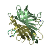
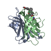
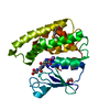
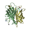
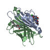
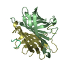
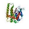
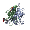
| Title | Spectroscopic and structural characterization of a genetically encoded direct sensor for protein-ligand interactions | ||||||
 Components Components | Streptavidin | ||||||
 Keywords Keywords | FLUORESCENT PROTEIN / unnatural amino acid / non-canonical amino acid / fluorescence / biosensor / small molecule biosensor / ligand detection | ||||||
| Function / homology |  Function and homology information Function and homology information | ||||||
| Biological species |  Streptomyces avidinii (bacteria) Streptomyces avidinii (bacteria) | ||||||
| Method |  X-RAY DIFFRACTION / SYNCHROTRON / MOLECULAR REPLACEMENT / Resolution: 1.502 Å X-RAY DIFFRACTION / SYNCHROTRON / MOLECULAR REPLACEMENT / Resolution: 1.502 Å | ||||||
 Authors Authors | Mills, J.H. / Gleason, P.R. / Simmons, C.R. / Henderson, J.N. / Kartchner, B.K. | ||||||
 Citation Citation | Journal: Biochemistry / Year: 2021 Title: Structural Origins of Altered Spectroscopic Properties upon Ligand Binding in Proteins Containing a Fluorescent Noncanonical Amino Acid. Authors: Gleason, P.R. / Kolbaba-Kartchner, B. / Henderson, J.N. / Stahl, E.P. / Simmons, C.R. / Mills, J.H. | ||||||
| History |
|
- Structure visualization
| Structure viewer | Molecule: MolmilJmol/JSmol |
|---|
- Downloads & links
-Download
| PDBx/mmCIF format | 6ud6.cif.gz | 71 KB | Display | PDBx/mmCIF format |
|---|---|---|---|---|
| PDB format | pdb6ud6.ent.gz | 51.6 KB | Display | PDB format |
| PDBx/mmJSON format | 6ud6.json.gz | Tree view | PDBx/mmJSON format | |
| Others |  Other downloads Other downloads |
-Validation report
| Arichive directory | https://data.pdbj.org/pub/pdb/validation_reports/ud/6ud6ftp://data.pdbj.org/pub/pdb/validation_reports/ud/6ud6 | HTTPS FTP |
|---|
-Related structure data
| Related structure data |  6uc3C  6ud1C  6udbC 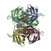 6udcC  3ry2S S: Starting model for refinement C: citing same article ( |
|---|---|
| Similar structure data |


-Links
 PDBj
PDBj- Assembly
| Deposited unit | 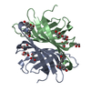
| ||||||||||||||||||
|---|---|---|---|---|---|---|---|---|---|---|---|---|---|---|---|---|---|---|---|
| 1 |
| ||||||||||||||||||
| 2 | 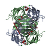
| ||||||||||||||||||
| Unit cell |
| ||||||||||||||||||
| Components on special symmetry positions |
|
-Components
| #1: Protein | Mass: 14542.706 Da / Num. of mol.: 2 Source method: isolated from a genetically manipulated source Source: (gene. exp.) Streptomyces avidinii (bacteria) / Production host: #2: Chemical | ChemComp-GOL /   Mass: 92.094 Da / Num. of mol.: 7 / Source method: obtained synthetically / Formula: C3H8O3 Mass: 92.094 Da / Num. of mol.: 7 / Source method: obtained synthetically / Formula: C3H8O3#3: Chemical |   Mass: 35.453 Da / Num. of mol.: 2 / Source method: obtained synthetically / Formula: Cl Mass: 35.453 Da / Num. of mol.: 2 / Source method: obtained synthetically / Formula: Cl#4: Water | ChemComp-HOH / |  Mass: 18.015 Da / Num. of mol.: 219 / Source method: isolated from a natural source / Formula: H2O Mass: 18.015 Da / Num. of mol.: 219 / Source method: isolated from a natural source / Formula: H2OHas ligand of interest | Y | Has protein modification | Y | |
|---|
-Experimental details
-Experiment
| Experiment | Method: X-RAY DIFFRACTION / Number of used crystals: 1 |
|---|
- Sample preparation
| Crystal | Density Matthews: 2.47 Å3/Da / Density % sol: 50.21 % |
|---|---|
| Crystal grow | Temperature: 298 K / Method: vapor diffusion, sitting drop / pH: 3.5 Details: 500 ul of 0.1 M citric acid, pH 3.5, 3M NaCl in the reservoir with 2 ul of reservoir buffer mixed with 2 ul of 10 mg/ml of protein in the drop. Crystals were soaked to pH 5.5 overnight with 3 exchanges |
-Data collection
| Diffraction | Mean temperature: 100 K / Serial crystal experiment: N |
|---|---|
| Diffraction source | Source: SYNCHROTRON / Site: ALS  / Beamline: 5.0.2 / Wavelength: 1 Å / Beamline: 5.0.2 / Wavelength: 1 Å |
| Detector | Type: DECTRIS PILATUS3 6M / Detector: PIXEL / Date: May 13, 2018 |
| Radiation | Protocol: SINGLE WAVELENGTH / Monochromatic (M) / Laue (L): M / Scattering type: x-ray |
| Radiation wavelength | Wavelength: 1 Å / Relative weight: 1 |
| Reflection | Resolution: 1.5→29.631 Å / Num. obs: 44561 / % possible obs: 99.97 % / Redundancy: 13.5 % / CC1/2: 1 / Net I/σ(I): 14 |
| Reflection shell | Resolution: 1.5→1.53 Å / Mean I/σ(I) obs: 2.1 / Num. unique obs: 2228 / CC1/2: 0.845 / % possible all: 100 |
- Processing
| Software |
| ||||||||||||||||||||||||
|---|---|---|---|---|---|---|---|---|---|---|---|---|---|---|---|---|---|---|---|---|---|---|---|---|---|
| Refinement | Method to determine structure: MOLECULAR REPLACEMENT Starting model: 3RY2 Resolution: 1.502→29.631 Å / SU ML: 0.11 / Cross valid method: THROUGHOUT / σ(F): 1.34 / Phase error: 19.32
| ||||||||||||||||||||||||
| Solvent computation | Shrinkage radii: 0.9 Å / VDW probe radii: 1.11 Å | ||||||||||||||||||||||||
| Displacement parameters | Biso max: 73.59 Å2 / Biso mean: 24.2635 Å2 / Biso min: 11.44 Å2 | ||||||||||||||||||||||||
| Refinement step | Cycle: final / Resolution: 1.502→29.631 Å
| ||||||||||||||||||||||||
| LS refinement shell | Resolution: 1.502→1.53 Å /
|
