 Movie
Movie Controller

[English] 日本語
 Yorodumi
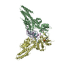
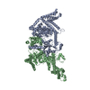
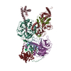
- PDB-6lml: Cryo-EM structure of the human glucagon receptor in complex with Gi1 -
+ Open data

- Basic information
| Entry | Database: PDB / ID: 6lml | |||||||||||||||||||||||||||
|---|---|---|---|---|---|---|---|---|---|---|---|---|---|---|---|---|---|---|---|---|---|---|---|---|---|---|---|---|
| Title | Cryo-EM structure of the human glucagon receptor in complex with Gi1 | |||||||||||||||||||||||||||
 Components Components |
| |||||||||||||||||||||||||||
 Keywords Keywords | SIGNALING PROTEIN / glucagon receptor / GPCR / Gi1 protein | |||||||||||||||||||||||||||
| Function / homology |  Function and homology information Function and homology informationglucagon receptor binding / regulation of glycogen metabolic process / glucagon receptor activity / : / feeding behavior / negative regulation of execution phase of apoptosis / response to starvation / positive regulation of calcium ion import / regulation of insulin secretion / peptide hormone binding ...glucagon receptor binding / regulation of glycogen metabolic process / glucagon receptor activity / : / feeding behavior / negative regulation of execution phase of apoptosis / response to starvation / positive regulation of calcium ion import / regulation of insulin secretion / peptide hormone binding / Synthesis, secretion, and deacylation of Ghrelin / adenylate cyclase inhibitor activity / positive regulation of protein localization to cell cortex / T cell migration / positive regulation of relaxation of smooth muscle / Adenylate cyclase inhibitory pathway / response to nutrient / cellular response to glucagon stimulus / D2 dopamine receptor binding / guanyl-nucleotide exchange factor activity / adenylate cyclase-inhibiting serotonin receptor signaling pathway / positive regulation of gluconeogenesis / G protein-coupled serotonin receptor binding / cellular response to forskolin / cellular response to starvation / positive regulation of insulin secretion involved in cellular response to glucose stimulus / response to activity / gluconeogenesis / regulation of mitotic spindle organization / chemokine-mediated signaling pathway / generation of precursor metabolites and energy / Regulation of insulin secretion / neuropeptide signaling pathway / response to prostaglandin E / hormone activity / positive regulation of cholesterol biosynthetic process / negative regulation of insulin secretion / G protein-coupled receptor binding / regulation of blood pressure / response to peptide hormone / centriolar satellite / G-protein beta/gamma-subunit complex binding / adenylate cyclase-modulating G protein-coupled receptor signaling pathway / positive regulation of insulin secretion / adenylate cyclase-inhibiting G protein-coupled receptor signaling pathway / Olfactory Signaling Pathway / Activation of the phototransduction cascade / G protein-coupled acetylcholine receptor signaling pathway / G beta:gamma signalling through PLC beta / Presynaptic function of Kainate receptors / Thromboxane signalling through TP receptor / Activation of G protein gated Potassium channels / Inhibition of voltage gated Ca2+ channels via Gbeta/gamma subunits / G-protein activation / Glucagon signaling in metabolic regulation / G beta:gamma signalling through CDC42 / Prostacyclin signalling through prostacyclin receptor / Synthesis, secretion, and inactivation of Glucagon-like Peptide-1 (GLP-1) / G beta:gamma signalling through BTK / photoreceptor disc membrane / ADP signalling through P2Y purinoceptor 12 / GDP binding / Glucagon-type ligand receptors / Sensory perception of sweet, bitter, and umami (glutamate) taste / Adrenaline,noradrenaline inhibits insulin secretion / Vasopressin regulates renal water homeostasis via Aquaporins / Glucagon-like Peptide-1 (GLP1) regulates insulin secretion / G alpha (z) signalling events / cellular response to catecholamine stimulus / ADP signalling through P2Y purinoceptor 1 / G beta:gamma signalling through PI3Kgamma / ADORA2B mediated anti-inflammatory cytokines production / adenylate cyclase-activating dopamine receptor signaling pathway / glucose homeostasis / Cooperation of PDCL (PhLP1) and TRiC/CCT in G-protein beta folding / GPER1 signaling / cellular response to prostaglandin E stimulus / heterotrimeric G-protein complex / G alpha (12/13) signalling events / Inactivation, recovery and regulation of the phototransduction cascade / G-protein beta-subunit binding / extracellular vesicle / sensory perception of taste / Thrombin signalling through proteinase activated receptors (PARs) / sperm principal piece / adenylate cyclase-activating G protein-coupled receptor signaling pathway / signaling receptor complex adaptor activity / retina development in camera-type eye / fibroblast proliferation / GTPase binding / G protein activity / midbody / secretory granule lumen / Ca2+ pathway / cell cortex / High laminar flow shear stress activates signaling by PIEZO1 and PECAM1:CDH5:KDR in endothelial cells / G alpha (i) signalling events / G alpha (s) signalling events / G alpha (q) signalling events / phospholipase C-activating G protein-coupled receptor signaling pathway Similarity search - Function | |||||||||||||||||||||||||||
| Biological species |  Homo sapiens (human) Homo sapiens (human) | |||||||||||||||||||||||||||
| Method | ELECTRON MICROSCOPY / single particle reconstruction / cryo EM / Resolution: 3.9 Å | |||||||||||||||||||||||||||
 Authors Authors | Qiao, A. / Han, S. / Li, X. / Sun, F. / Zhao, Q. / Wu, B. | |||||||||||||||||||||||||||
| Funding support |  China, 8items China, 8items
| |||||||||||||||||||||||||||
 Citation Citation | Journal: Science / Year: 2020 Title: Structural basis of G and G recognition by the human glucagon receptor. Authors: Anna Qiao / Shuo Han / Xinmei Li / Zhixin Li / Peishen Zhao / Antao Dai / Rulve Chang / Linhua Tai / Qiuxiang Tan / Xiaojing Chu / Limin Ma / Thor Seneca Thorsen / Steffen Reedtz-Runge / ...Authors: Anna Qiao / Shuo Han / Xinmei Li / Zhixin Li / Peishen Zhao / Antao Dai / Rulve Chang / Linhua Tai / Qiuxiang Tan / Xiaojing Chu / Limin Ma / Thor Seneca Thorsen / Steffen Reedtz-Runge / Dehua Yang / Ming-Wei Wang / Patrick M Sexton / Denise Wootten / Fei Sun / Qiang Zhao / Beili Wu /   Abstract: Class B G protein-coupled receptors, an important class of therapeutic targets, signal mainly through the G class of heterotrimeric G proteins, although they do display some promiscuity in G protein ...Class B G protein-coupled receptors, an important class of therapeutic targets, signal mainly through the G class of heterotrimeric G proteins, although they do display some promiscuity in G protein binding. Using cryo-electron microscopy, we determined the structures of the human glucagon receptor (GCGR) bound to glucagon and distinct classes of heterotrimeric G proteins, G or G These two structures adopt a similar open binding cavity to accommodate G and G The G binding selectivity of GCGR is explained by a larger interaction interface, but there are specific interactions that affect G more than G binding. Conformational differences in the receptor intracellular loops were found to be key selectivity determinants. These distinctions in transducer engagement were supported by mutagenesis and functional studies. | |||||||||||||||||||||||||||
| History |
|
- Structure visualization
| Movie |
Movie viewer |
|---|---|
| Structure viewer | Molecule: MolmilJmol/JSmol |
- Downloads & links
-Download
| PDBx/mmCIF format | 6lml.cif.gz | 232 KB | Display | PDBx/mmCIF format |
|---|---|---|---|---|
| PDB format | pdb6lml.ent.gz | 175.5 KB | Display | PDB format |
| PDBx/mmJSON format | 6lml.json.gz | Tree view | PDBx/mmJSON format | |
| Others |  Other downloads Other downloads |
-Validation report
| Arichive directory | https://data.pdbj.org/pub/pdb/validation_reports/lm/6lmlftp://data.pdbj.org/pub/pdb/validation_reports/lm/6lml | HTTPS FTP |
|---|
-Related structure data
| Related structure data | 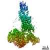 0918MC 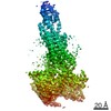 0917C  6lmkC M: map data used to model this data C: citing same article ( |
|---|---|
| Similar structure data |


-Links
 PDBj
PDBj


- Assembly
| Deposited unit | 
|
|---|---|
| 1 |
|
-Components
-Guanine nucleotide-binding protein ... , 3 types, 3 molecules ABC
| #1: Protein | Mass: 40414.047 Da / Num. of mol.: 1 / Mutation: S47N,G203A,E245A,A326S Source method: isolated from a genetically manipulated source Source: (gene. exp.) Homo sapiens (human) / Gene: GNAI1 / Production host:  unidentified baculovirus / References: UniProt: P63096 unidentified baculovirus / References: UniProt: P63096 |
|---|---|
| #2: Protein | Mass: 38744.371 Da / Num. of mol.: 1 Source method: isolated from a genetically manipulated source Source: (gene. exp.) Homo sapiens (human) / Gene: GNB1 / Production host: unidentified baculovirus / References: UniProt: P62873 |
| #3: Protein | Mass: 7861.143 Da / Num. of mol.: 1 Source method: isolated from a genetically manipulated source Source: (gene. exp.) Homo sapiens (human) / Gene: GNG2 / Production host: unidentified baculovirus / References: UniProt: P59768 |
-Antibody / Protein/peptide / Protein , 3 types, 3 molecules DER
| #4: Antibody | Mass: 26337.307 Da / Num. of mol.: 1 Source method: isolated from a genetically manipulated source Source: (gene. exp.) unidentified baculovirus |
|---|---|
| #5: Protein/peptide | Mass: 3486.781 Da / Num. of mol.: 1 / Source method: obtained synthetically / Source: (synth.) Homo sapiens (human) / References: UniProt: P01275 |
| #6: Protein | Mass: 48571.410 Da / Num. of mol.: 1 / Mutation: E126R,T200W,A366M Source method: isolated from a genetically manipulated source Source: (gene. exp.) Homo sapiens (human) / Gene: GCGR / Production host: unidentified baculovirus / References: UniProt: P47871 |
-Details
| Has protein modification | Y |
|---|
-Experimental details
-Experiment
| Experiment | Method: ELECTRON MICROSCOPY |
|---|---|
| EM experiment | Aggregation state: PARTICLE / 3D reconstruction method: single particle reconstruction |
- Sample preparation
| Component | Name: Complex of glucagon receptor bound to glucagon, Gi1 protein and antibody Type: COMPLEX / Entity ID: all / Source: RECOMBINANT |
|---|---|
| Source (natural) | Organism: Homo sapiens (human) |
| Source (recombinant) | Organism: unidentified baculovirus |
| Buffer solution | pH: 7.5 |
| Specimen | Embedding applied: NO / Shadowing applied: NO / Staining applied: NO / Vitrification applied: YES |
| Vitrification | Cryogen name: ETHANE |
- Electron microscopy imaging
| Experimental equipment |  Model: Titan Krios / Image courtesy: FEI Company |
|---|---|
| Microscopy | Model: FEI TITAN KRIOS |
| Electron gun | Electron source:  FIELD EMISSION GUN / Accelerating voltage: 300 kV / Illumination mode: FLOOD BEAM FIELD EMISSION GUN / Accelerating voltage: 300 kV / Illumination mode: FLOOD BEAM |
| Electron lens | Mode: BRIGHT FIELD |
| Image recording | Electron dose: 1.875 e/Å2 / Film or detector model: GATAN K2 SUMMIT (4k x 4k) |
- Processing
| CTF correction | Type: PHASE FLIPPING AND AMPLITUDE CORRECTION | ||||||||||||||||||||||||
|---|---|---|---|---|---|---|---|---|---|---|---|---|---|---|---|---|---|---|---|---|---|---|---|---|---|
| Symmetry | Point symmetry: C1 (asymmetric) | ||||||||||||||||||||||||
| 3D reconstruction | Resolution: 3.9 Å / Resolution method: FSC 0.143 CUT-OFF / Num. of particles: 312974 / Symmetry type: POINT | ||||||||||||||||||||||||
| Refinement | Stereochemistry target values: GeoStd + Monomer Library + CDL v1.2 | ||||||||||||||||||||||||
| Displacement parameters | Biso mean: 75.59 Å2 | ||||||||||||||||||||||||
| Refine LS restraints |
|
